

A Vestibular Detective Story Highlighting Sequential Nystagmus, Inhibitory Mechanisms, and Clinical Reasoning Under Uncertainty


Textbook BPPV cases are satisfying, but the complex ones truly forge our clinical skills. They push us to observe more keenly, question our assumptions, revisit fundamentals, and sometimes navigate significant diagnostic uncertainty over multiple visits. This article chronicles one such case – a three-visit journey involving conflicting symptoms and signs, perplexing nystagmus, ruled-out hypotheses, concerns about central pathology, and ultimately, a breakthrough achieved by returning to basic principles and performing a 'diagnosis of exclusion' test. Revisiting fundamentals is not a sign of weakness but a testament to our commitment to thorough and accurate diagnosis.
Visit 1: Right-Sided Complaint, Left-Sided Surprise, Lingering Question
A patient presented reporting recurrent vertigo, emphasizing symptoms that occurred primarily when lying on their right side. Standard practice often involves checking the contralateral, asymptomatic side first, so I initiated testing with the Left Dix-Hallpike.
The result was immediate and twofold:
Act I – Clear Left BPPV: A crisp, upbeat nystagmus with left torsion appeared instantly, fatiguing classically after a short duration. This confirmed the Left Posterior Semicircular Canal (PSC) and BPPV (Canalithiasis). The patient expressed surprise, having never noted issues on this side.
Act II – The Persistent Puzzle: As soon as the upbeat nystagmus subsided while still in the Left Dix-Hallpike position, a second pattern emerged: persistent, non-fatiguing, downbeating nystagmus with left torsion.
My immediate thought process grappled with Act II:
Initial Hypothesis: Could debris move backward (ampullopetally) in the Left PSC I just saw active?
Rapid Self-Correction: No, that did not fit. Ampullopetal movement/jamming in the Left PSC should cause downbeat torsion to the RIGHT. The observed LEFT torsion meant this hypothesis was incorrect. The vector was wrong.
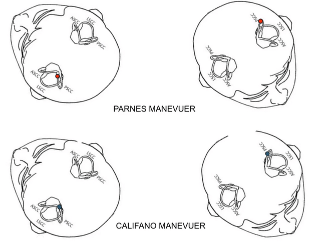
I treated the identified Left PSC BPPV from Act I with a Parnes maneuver - AKA: Lying down Semont (details of specific maneuvers less critical here than the fact it was addressed).

However, the persistent downbeat/left torsional nystagmus from Act II remained even after the Left-sided treatment concluded (while the patient was still in a dependent position), only resolving upon sitting up. This strongly suggested its origin was separate from the Left PSC canalithiasis. I was left with a question mark about this persistent pattern.
Visit 2: Deepening Mystery, Ruling Out Possibilities
The patient returned. Reassessment revealed that the Left PSC BPPV had recurred (or was incompletely treated). I addressed the Left PSC BPPV again. The primary focus shifted to deciphering the persistent downbeat nystagmus and left torsion seen during the Left Dix-Hallpike.
Frankly, I was wrestling with confidently differentiating the torsion direction in this complex, persistent pattern. Was it indeed left, or could I be misinterpreting it as correct? This uncertainty fueled my next steps, attempting to rule out possibilities based on both potential torsion directions:
Testing Hypothesis: Downbeat/RIGHT Torsion (L PSC Ampullopetal)? If I was mistaken and the torsion was right, it could imply debris moving ampullopetally or adhering to the cupula in the Left PSC. I performed a Quick Liberatory Maneuver (Califano technique) (functionally similar to Parnes but with a twist ) targeting Left PSC cupulolithiasis/ampullopetal debris to test this.
***Crucial Difference: With a Parnes, Epley, or Semont, you pause to ensure the debris reaches the most dependent portion of the canal - this could take several seconds with a hold time of a recommended 30 to two minutes. In the Califano (Quick Liberatory Maneuver), you must move IMMEDIATELY as you want the debris to move AWAY FROM the ampulla. It starts with where the debris is in the canal - The anterior arm (Parnes) versus the posterior arm (Califano).
Result: Negative. There was no change and no provocation of nystagmus that would suggest clearing the canal in this way. This made a Left PSC source for the persistent nystagmus highly unlikely.
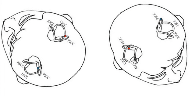
Testing Hypothesis: Downbeat/LEFT Torsion (L AC Involvement?) If the torsion was definitively left, the Left Anterior Canal (AC) is a prime suspect for downbeating nystagmus. To test this, I performed a Modified Yacovino maneuver (Bhandari, 2022) targeting the Left AC.
Result: Negative. There was no therapeutic effect and no expected nystagmus when sitting up. This made Left AC involvement unlikely.
I had now systematically tested and found negative results for the most likely Left-sided explanations (PSC ampullopetal/cupulo and AC) for the persistent downbeat nystagmus, regardless of my lingering uncertainty about the subtle torsion. The patient was sent home, the puzzle unsolved.
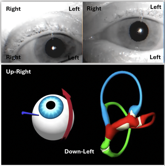
Author's Note & Clinical Pearl: Accurately judging torsional components, especially in persistent or low-amplitude nystagmus, can be challenging. This is where high-quality video goggles are invaluable. For readers, understanding the difference is key. I strongly recommend viewing examples comparing these vectors.

Visit 3: The Central Scare, The "Stop and Think," The Breakthrough
The patient returned for the third visit. The Left Dix-Hallpike still provoked that persistent, non-fatiguing, downbeat nystagmus with what I now felt more confident about: LEFT torsion. The persistence, the unusual vector, and the lack of response to peripheral maneuvers significantly raised my concern.
Considering Central Positional Vertigo: Was I missing a central cause? Persistent downbeating nystagmus can be a red flag, especially if truly direction-fixed. I had a frank discussion with the patient about the possibility that this might not be typical BPPV but potentially a central positional vertigo syndrome requiring further neurological workup.
The 'Stop and Think' Moment: Before referring out, I paused. "Stop, Brian," I told myself. "Think fundamentally." This moment, which I call the 'Stop and Think' moment, is a crucial part of the diagnostic process. It is a reminder to step back, reevaluate the situation, and consider all possible explanations. What peripherally causes persistent downbeat/left torsion? We had effectively ruled out the Left AC. The only other primary peripheral candidate was inhibition of the Right Posterior Canal. Solidifying the Hypothesis: Could the Right PSC be inhibited during a Left Dix-Hallpike? It seemed counterintuitive, given the expected nullification. But what if her anatomy was slightly "wonky"? What if that specific Left DHP position placed her Right PSC at an angle, allowing debris to move ampullopetally, causing a sustained, cupulolithiasis-like inhibitory pressure on the ampulla? It was the only remaining peripheral explanation that fit the observed nystagmus vector.
The Diagnosis of Exclusion Test: I decided, "What do I have to lose?" If Right PSC inhibition during L DHP was happening, then direct testing of the Right PSC should reveal its excitatory response if canalithiasis was present. My next step became clear: perform a Right Dix-Hallpike. This test, known as the 'diagnosis of exclusion' test, involves ruling out all other possible diagnoses before concluding the correct one. In this case, it was used to rule out other potential causes of the persistent nystagmus, leading to the proper diagnosis.
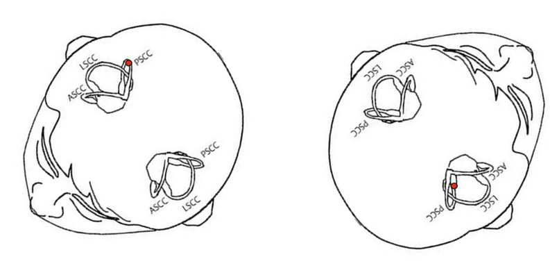
The Confirmation ("Aha!"): I performed the Right Dix-Hallpike. The immediate and dramatic result: severe, unambiguous, upbeat nystagmus with right torsion – classic, active Right PSC BPPV (Canalithiasis). i performed the Parnes Manevuer for the right, and the vertigo and symptoms resolved. The mystery of the persistent downbeat/left torsion was solved: it was the inhibitory signature of this underlying Right PSC issue, manifesting only during the contralateral test. The 'inhibitory signature' concept refers to the unique nystagmus pattern that can indicate the presence of a specific vestibular disorder. Once we understood its inhibitory nature, the persistent downbeat/left torsion was a clear sign of the underlying Right PSC issue.
The Reflection: Why the Delay? (Parnes vs. Epley)
In retrospect, why wasn't this Right PSC BPPV caught earlier? My consistent use of the Parnes maneuver to treat the Left PSC on visits 1 and 2 might have played a role. By skipping the standard second position of the Epley maneuver (head 45° right, supine), I potentially missed a diagnostic window where the excitatory response of the Right PSC might have been revealed during the treatment for the left side. This was not a treatment error for the left but a reflection of how maneuver choice can impact incidental findings.
The Resolution: Finally Treating the Root Cause
I performed the Parnes maneuver for the Right PSC with the diagnosis clear. Moving the patient from the provoking Right Dix-Hallpike position towards the final nose-down-on-the-left position, she experienced another severe upbeat/right torsion, confirming successful debris migration through the canal. After resting, she sat up. Reassessment showed complete resolution – negative tests bilaterally, no more vertigo.
Clinical Takeaways from a Diagnostic Odyssey:
This multi-visit journey underscored several invaluable lessons:
Trust, But Verify (Symptoms vs. Signs): Listen to the patient's primary complaint, even if initial tests point elsewhere. It often holds clues to the underlying issue.
Embrace Uncertainty, Test Systematically: It is okay to be unsure, especially with subtle findings like torsion. Use systematic testing of different hypotheses (L PSC ampullo/cupulo, L AC, R PSC inhibition) to narrow possibilities.
Know Nystagmus Vectors (Excitatory & Inhibitory): Deep knowledge of expected nystagmus for excitation and inhibition of each canal is non-negotiable for solving complex cases.
Consider Central, But Exhaust Peripheral: Persistent atypical nystagmus warrants considering central causes, but thoroughly explore and rule out complex peripheral explanations first.
Fundamentals First: When stuck, return to basic principles: What structures cause this specific nystagmus vector?
Reflect on Process: Consider how testing order and maneuver choices might influence findings or potentially create diagnostic "blind spots."
Conclusion: Celebrating the Process
This case was a powerful reminder that vestibular diagnostics can be a true detective story, complete with twists, red herrings, and moments of uncertainty. The path to the correct diagnosis was not linear but involved careful observation, hypothesis testing, self-correction, revisiting fundamental principles, and ultimately trusting the exclusion process. By sharing these complex journeys, we collectively enhance our ability to recognize, interpret, and successfully manage the intricate presentations our patients bring to us.