


Active vs. Passive Headshake Testing in Vestibular Assessment: Unveiling the Nuances

The headshake test (HST) is a cornerstone in assessing vestibular disorders, providing crucial insights into the integrity of the vestibulo-ocular reflex (VOR), a fundamental mechanism for maintaining stable vision during head movements. However, the choice between active (patient-initiated) and passive (clinician-initiated) head shaking has sparked an ongoing and engaging debate among clinicians. Each approach potentially influences the test's outcomes and interpretation, making it a topic of significant interest and discussion. This article aims to delve into the complexities of both methods, exploring the underlying physiological mechanisms, the potential impact of cervical and vestibulo-collic reflexes, the available evidence, and practical guidelines for administering the test.
Background: The Vestibular System and Head Movements
The vestibular system, nestled within the inner ear, is pivotal in maintaining balance and coordinating head and eye movements. The VOR, an essential reflex arc within this system, ensures that our eyes remain fixed on a target even as our head moves. When the vestibular system is compromised, as seen in various vestibulopathies, this reflex can become impaired, leading to symptoms like dizziness, vertigo, and nystagmus (involuntary eye movements).
The HST capitalizes on this relationship by inducing brief head movements, which can provoke nystagmus in individuals with vestibular dysfunction. However, how these head movements are initiated—actively by the patient or passively by the clinician—can introduce subtle yet potentially significant differences in the elicited responses.
Active vs. Passive Head Shake: Physiological Considerations
Active Head Shake: When patients actively shake their heads, they engage the vestibular system and the neck proprioceptors. These sensory receptors provide feedback about head and neck position, contributing to reflexes like the vestibulo-collic reflex (VCR) and cervico-collic reflex (CCR). The VCR stabilizes the head in response to vestibular input, while the CCR stabilizes the neck in response to neck movement. During active head shaking, activating these reflexes may introduce compensatory mechanisms that could mask subtle vestibular deficits, underscoring their significant role in the headshake test.
Passive Head Shake: In contrast, when the clinician passively moves the patient's head, the influence of neck proprioceptors might be minimized. This could potentially isolate vestibular responses, allowing for a "purer" assessment of the VOR and making it easier to detect subtle abnormalities. However, it's essential to consider that dysfunction in the cervical proprioceptors or the VCR may lead to an increased nystagmus response during headshake testing. This is because these systems, when functioning correctly, help to dampen the VOR response. When impaired, they may no longer provide this dampening effect, leading to a more pronounced nystagmus.
Evidence from the Literature
While research on active versus passive head shaking in the HST is ongoing, several studies have shed light on the potential differences between these approaches:
MacDougall et al. (2007): This study found that while the frequency of nystagmus was similar between active and passive head shaking, the type of nystagmus varied with age in patients with vestibular neuritis. Younger patients exhibited more nystagmus with active head shaking, while older patients showed more with passive head shaking.
Tusa et al. (2004): This research revealed that a small percentage of healthy individuals exhibit head-shaking nystagmus (HSN), suggesting that HSN alone might not be a definitive indicator of vestibular dysfunction.
Kilicarslan et al. (2018): This study focused on the effectiveness of active head shaking in diagnosing vestibular neuritis, finding it to be a valuable tool in clinical assessment.
Sralab Rehab Measures Database: This comprehensive resource recommends passive head shaking by the clinician but does not explicitly address the differences between the two approaches.
How to Perform the Headshake Test
Regardless of whether you choose active or passive headshake testing, the following steps should be followed:
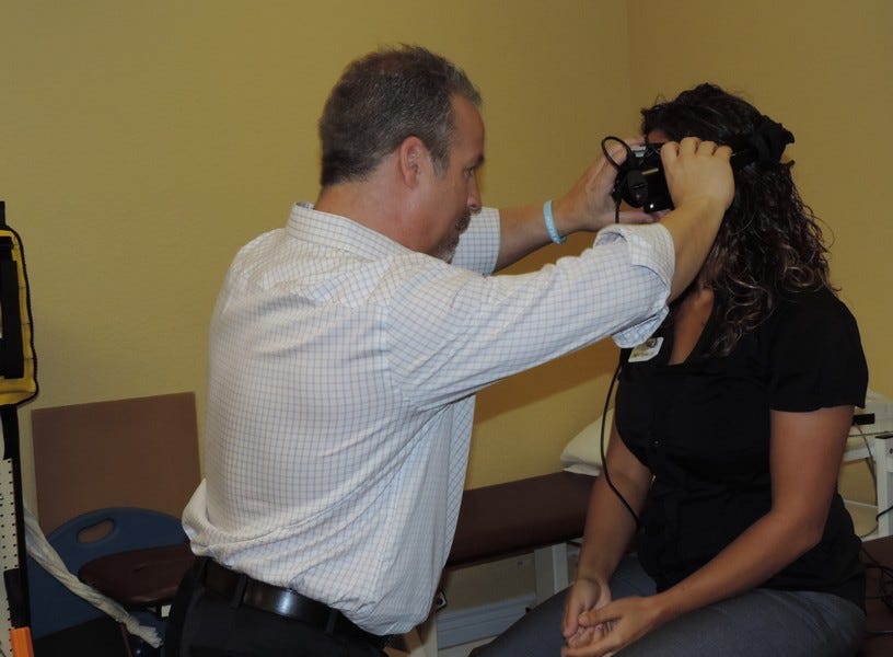
Patient Preparation: Ensure the patient is seated comfortably with their head and neck relaxed. If using video nystagmography (VNG), place the goggles on the patient and ensure proper calibration.
Head Positioning: Tilt the patient's head forward approximately 30 degrees to align the horizontal semicircular canals with the plane of movement.
Head Movement:
Active: Instruct the patient to shake their head horizontally (as if saying "no") at a comfortable pace for 20-30 seconds, even up to one minute. Please encourage them to maintain a consistent speed and amplitude.
Passive: Gently grasp the patient's head and oscillate it horizontally at a frequency of 2-3 Hz (120-180 beats per minute) for a minimum of 20 seconds, up to 1 minute if necessary.
Observation: After the head shaking stops, carefully observe the patient's eyes for the presence of nystagmus. Note the direction, amplitude, and duration of any nystagmus. If using VNG, the nystagmus will be recorded for later analysis.
Clinical Implications and Best Practices
Given these findings, clinicians should carefully consider the following when choosing between active and passive head shake testing:
Patient Factors: Age, underlying medical conditions (particularly those affecting the cervical spine), and the specific type of suspected vestibular disorder can influence the head-shaking method.
Test Sensitivity: The sensitivity of active versus passive head shaking in detecting specific vestibular pathologies remains an area that requires further research. Until then, clinicians should be mindful of the potential for both false positives and negatives with either method, particularly in cases where the cervical or vestibule-collect function may be compromised. This underscores the need for continued investigation into the most effective use of the headshake test.
Patient Comfort and Tolerance: To minimize discomfort and ensure feasibility, passive head shaking may be preferred for patients experiencing acute vestibular symptoms or those with physical limitations.
Horizontal vs. Vertical Headshake Testing: While horizontal headshake testing remains the standard approach, clinicians may consider vertical headshake testing in specific cases, such as suspected vertical canal dysfunction. However, the interpretation of vertical nystagmus can be complex due to the simultaneous stimulation of multiple semicircular canals.
Conclusion
The headshake test is a valuable tool in the assessment of vestibular function. By understanding the nuances of active and passive head shaking, considering individual patient factors, and adhering to standardized protocols, clinicians can optimize the diagnostic utility of this test and ultimately improve outcomes for individuals with vestibular disorders.
References & Further Readings
MacDougall, H. G., McGarvie, A., & Halmagyi, G. M. (2007). Active versus passive head-shaking nystagmus. Journal of Neurology, Neurosurgery & Psychiatry, 78(4), 354-359.
Tusa, R. J., Kaplan, P. W., Hain, T. C., & Herdman, S. J. (2004). Head-shaking nystagmus in healthy subjects. Otology & Neurotology, 25(2), 145-148.
Kilicarslan, S., Yilmazer, C., & Yilmaz, O. (2018). Examination of the effectiveness of the Active Head Shaking Nystagmus Test for the diagnosis of vestibular neuritis. Journal of International Advanced Otology, 14(3), 351-355.
Sralab Rehab Measures Database. The Head-Shaking Nystagmus Test. Available from: https://www.sralab.org/rehabilitation-measures