
Beyond the Textbook: When BPPV Plays Hard to Get - Reviewing Case Studies


As a physical therapist specializing in vestibular and balance disorders, I often see patients whose symptoms don't fit neatly into a diagnostic box. Benign Paroxysmal Positional Vertigo (BPPV) is typically straightforward, but there are always those head-scratching cases. A recent 2024 article, ‘Case Report: Keep your eyes open! Nystagmus guides atypical BPPV’ by Ludwig and Schubert truly resonates with the challenges we face in the clinic. It's a powerful reminder that while protocols are vital, keen observation and a deep understanding of vestibular physiology are our most potent diagnostic tools.
This article highlights four unique BPPV presentations that underscore a critical message: always trust the nystagmus. The specific eye movements reveal the truth even when the head position seems to tell one story. Let's dive into these fascinating cases.
Case 1: Bilateral Dix-Hallpike Nystagmus, Unilateral Problem
Imagine a patient, a 42-year-old woman with a history of BPPV, reporting vertigo when rolling to the left in bed or lying supine. We perform a Dix-Hallpike Test (DHT). Surprisingly, the right and left DHTs elicit upbeat and left torsional nystagmus. A glance might suggest bilateral posterior canal BPPV, a rare occurrence.
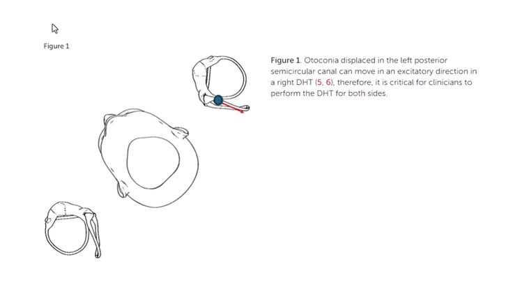
However, a closer look at the nystagmus tells a different story. The left torsional component remained consistent across both tests, and we could accentuate it by having the patient change her gaze. This consistency pointed directly to a left posterior semicircular canal involvement. Furthermore, the nystagmus and vertigo were significantly more intense during the Left DHT, where the affected left posterior canal is most dependent. The nystagmus also persisted for an extended duration, beyond 40-60 seconds, which raised a red flag for cupulolithiasis.
This case taught us to look beyond just the presence of nystagmus in a test.
'The specific direction of the torsional component, regardless of the head position, coupled with intensity differences, was the true guide.'
This patient likely had left posterior canal cupulolithiasis, with otoconia adhering to the cupula, explaining the persistent nystagmus and initial resistance to an Epley maneuver. She eventually found success with a Semont-plus maneuver.
Brian Werner Note: Another possible phenomenon could be that the debris moves through the common crus into the posterior arm of the anterior canal and the test side. This would cause inhibition to the ASC and create an upbeat torsion to the right. You would not see the upbeat torsion on the right side with the Dix-Hallpike test.
Case 2: The Dance of Excitatory and Inhibitory Nystagmus
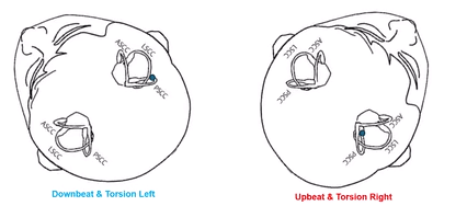
Next, consider a 44-year-old woman with a month-long history of episodic vertigo. In her Left DHT, we observed downbeat and left torsional nystagmus, lasting over 60 seconds (basically sitting on the cupula), yet she reported only generalized dizziness, not true vertigo. Then, a robust, upbeat, right torsional nystagmus appeared in the right DHT, consistent with a right posterior semicircular canalithiasis.
This case showcases the complexity of vestibular responses. The downbeat and left torsional nystagmus in the Left DHT wasn't an anomaly but an inhibitory response (ampullopetal flow) from the opposite right posterior semicircular canal. How? In that specific position, the otoconia in the right posterior canal moved in a direction that inhibited the ampulla, causing an opposite nystagmus direction to what we typically expect from excitation. This distinct pattern and the excitatory nystagmus in the right DHT solidified their diagnosis of a unilateral right posterior canal BPPV. The patient found relief with an Epley maneuver.
Case 3: The Surprise Nystagmus During Supine Roll Testing
An 80-year-old man, with multiple health issues and limited neck range of motion, presented for positional vertigo. He had a history of successfully treated right posterior canal BPPV. When we positioned him from long sitting to supine with his head flexed 30 degrees (a position we use to prepare for the Supine Roll Test for horizontal canal BPPV), something unexpected happened. After a brief latency, he developed upbeat and right torsional nystagmus.
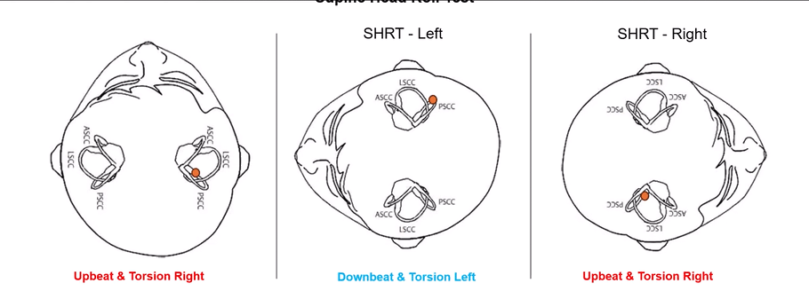
This was a critical moment. Despite being in a ‘horizontal canal’ testing position, the nystagmus was vertical-torsional, indicating a posterior canal issue. Going through the Supine Head Roll Test to the left, we could see that the patient presented with a downbeat and torsional left nystagmus that fatigued (what could this be: Left anterior canal or right posterior canal). Rolling the patient to the right side, the patient presented with a classic upbeat and torsional nystagmus. His past medical history further supported this. Recognizing this, we swiftly moved to a Right Epley Maneuver, confirming the right posterior canal BPPV and successfully treated.
This early identification was invaluable, especially for an elderly patient with musculoskeletal limitations, as it reduced unnecessary positional changes. It highlights how vital it is to identify canal-specific nystagmus patterns independently of the positional test.
Case 4: The Multi-Canal Maze
The final case involves a 74-year-old man with a two-week history of positional vertigo and gait unsteadiness. This complex, multi-visit scenario tested diagnostic acumen, as he presented with a mix of nystagmus types across various tests.
At one point, during a Left Supine Roll Test, he initially showed a right-beating apogeotropic nystagmus (pointing to horizontal canal BPPV) which then transitioned to an upbeat and left torsional nystagmus (indicating posterior canal BPPV). This simultaneous presentation clearly showed multi-canal BPPV, affecting both his horizontal and posterior canals. Further testing, including bow and lean tests, helped us isolate the specific affected canals.
This patient's journey emphasized that otoconia can migrate, and sometimes, more than one canal is involved. We also learned that nystagmus can reverse direction upon returning to sitting, which, surprisingly, helped rule out anterior canal BPPV in his case. Ultimately, successful treatment required addressing both the horizontal and posterior canal issues.
The Takeaway for Clinicians
These cases from Ludwig and Schubert serve as a potent reminder for all vestibular clinicians, including myself, Brian Werner, PT, MPT. BPPV might be common, but its presentations are not always textbook perfect.
You must understand that each canal has an excitatory nystagmus and an inhibitory nystagmus. Also, each canal has a mirrored canal that will produce the same nystagmus.
Your eyes are your best diagnostic tool. Obsessively observe the direction, intensity, and duration of nystagmus.
Don't rely solely on the test position. The nystagmus itself often holds the key to the specific canal and type of BPPV.
Use technology wisely. Using fixation-removed testing (VestibularFirst/Vesticam) is critical, as visual fixation can mask significant nystagmus.
Understand vestibular physiology deeply. The underlying mechanics of how each canal responds to otoconial displacement will guide you through atypical scenarios.
Be prepared for anything. Multi-canal BPPV exists, and presentations can be fluid.
In the complex world of balance disorders, we sometimes need to ‘fly by the seat of our pants,’ relying on our instincts and experience when the path isn't clear. But with a solid foundation in vestibular physiology and meticulous observation, we can successfully navigate even the most atypical BPPV cases and get our patients back to balance.