
⚕️ Decoding the Dark: Pupillary Unrest as an Objective Vestibular Marker (Updated: 12/12/2025)

The Vestibular Clinician’s View Through the Infrared Goggles
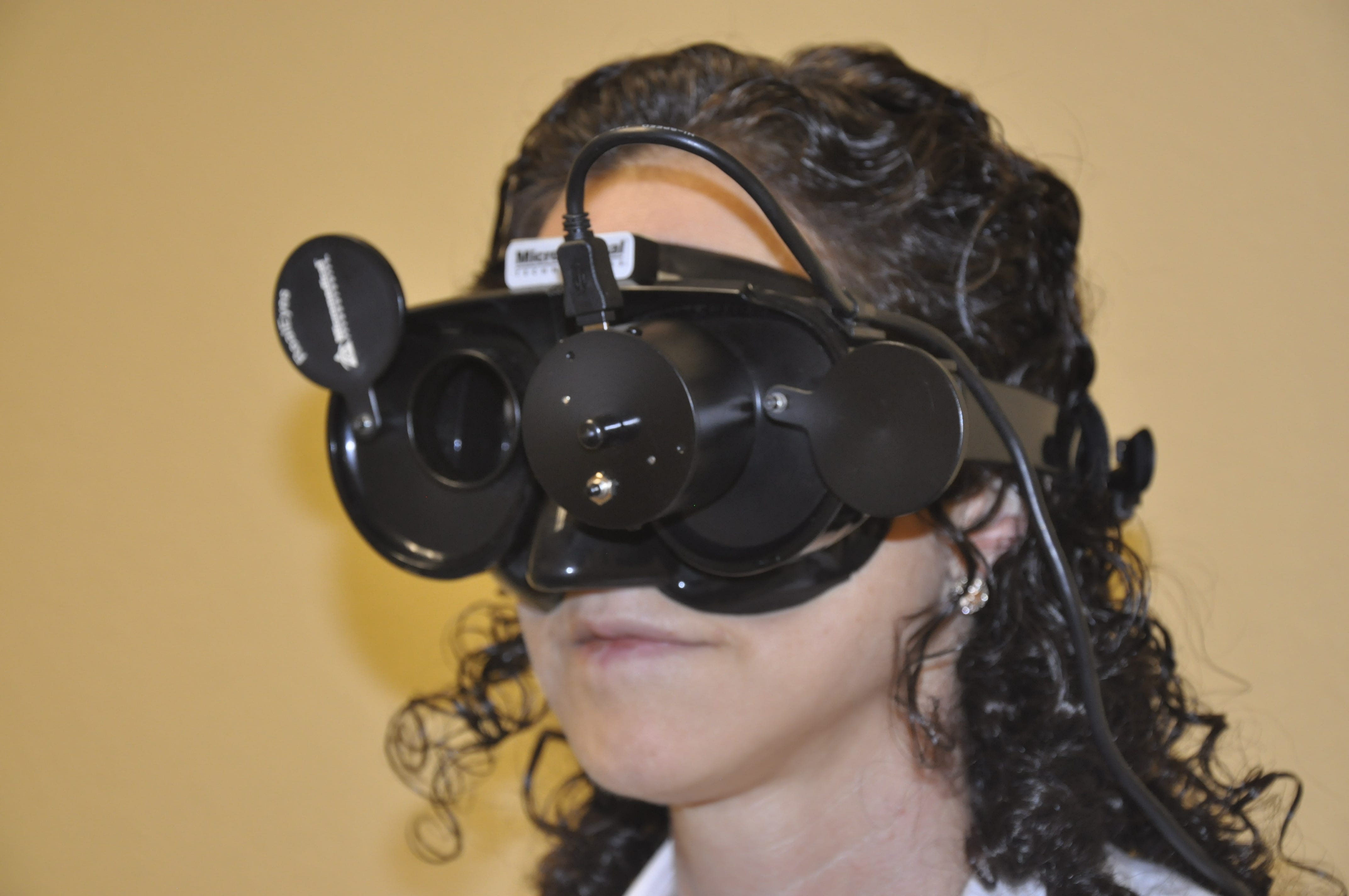
For the vestibular professional, the darkness of the infrared video goggle test is a moment of clarity. We search for subtle yet definitive eye movements—nystagmus (rapid, rhythmic eye movements)—that indicate inner-ear dysfunction. What about the spontaneous, erratic pupil fluctuation? This dilation and constriction, especially in the dark, is called Pupillary Unrest or Hippus (involuntary, rhythmic changes in pupil size).
Often dismissed as benign drowsiness in healthy individuals, research suggests that the ‘dancing pupil’ (pupillary hippus) offers a valuable, objective window into a patient’s autonomic state. This finding is particularly significant in neurological disorders, given that autonomic dysfunction (dysautonomia) is a common feature of Post-Concussion Syndrome (PCS). Recent studies have also identified this phenomenon as a potential clinical hallmark of specific vestibular pathologies, most notably Vestibular Migraine (Gufoni et al., 2023).
However, this pupillary unrest appears to reflect broader central autonomic dysregulation rather than a single disease. Consequently, it may also be present in other neurological conditions, including acute stroke. To fully distinguish among these potential causes, we must examine the specific underlying autonomic mechanisms in each pathology.
The Autonomic Tug-of-War: The Origin of Unrest
The pupil’s size is regulated by a delicate, continuous battle between the two branches of the Autonomic Nervous System (ANS), which controls involuntary body functions:
The Parasympathetic System (PNS): This system, known as the ‘rest and digest’ controller, drives the iris sphincter muscle, causing the pupil to constrict, a process called miosis (pupil shrinking).
The Sympathetic System (SNS): The ‘fight or flight’ system contracts the iris dilator muscle, causing the pupil to dilate (mydriasis).
Pupillary Unrest in the dark, and particularly the slow, high-amplitude oscillations often called ‘fatigue waves,’ reflects an instability in the central parasympathetic input to the Edinger-Westphal nucleus (a brain region controlling pupil size). When a patient is under-aroused or drowsy, this central control fluctuates, leading to the characteristic, repetitive, slow oscillations of constriction and dilation.
‘Pupillary Unrest serves as a real-time, objective marker of autonomic nervous system instability and reduced arousal.’
The Headshake Test and Post-Stimulation Pupillary Unrest
A critical observation encountered in the clinic involves the 20-second vigorous Headshake Test. This maneuver, designed to maximize the neural activity of the semicircular canals, sometimes yields an immediate and profound development of Hippus upon cessation. This finding moves the phenomenon beyond simple drowsiness and places it firmly within the context of vestibular-autonomic interplay.
The Headshake Test achieves two primary outcomes:
Neural Imbalance: In a patient with an underlying unilateral weakness, the 20 seconds of high-velocity shaking maximizes the velocity storage mechanism, creating a significant and temporary difference in neural firing between the two sides. When the shaking stops, the rebound elicits a strong, transient headshake nystagmus.
Massive Neural Load: The maneuver introduces a substantial neural load and stress into the entire vestibular network, which projects extensively to the brainstem’s autonomic control centers.
The immediate appearance of Pupillary Unrest (Hippus) after this intense vestibular stimulation suggests that the vestibular input itself—the profound temporary imbalance and resulting post-stimulatory neural stress—is directly destabilizing the autonomic control centers that regulate the pupil.
The mechanism points toward:
Autonomic Overload: The sudden cessation of the high-velocity vestibular input, combined with the maximal nystagmus that follows, may temporarily overload the central vestibular nuclei and the adjacent centers of the ANS, leading to a breakdown in the fine-tuned, dual control of the pupil.
Central Sensitization: In patients with conditions such as Vestibular Migraine (VM) or Persistent Postural-Perceptual Dizziness (PPPD), in which the central nervous system is hypothesized to be hyperresponsive, this intense vestibular stimulus acts as a trigger, revealing underlying autonomic lability through an exaggerated pupillary response.
The Vestibular Connection
The vestibular system is not solely an organ of balance and eye movement; it is intimately linked with the entire autonomic nervous system. (Physiological evidence that the vestibular system participates in autonomic and respiratory control, 2025) Vestibular inputs project extensively to brainstem nuclei that regulate autonomic functions such as blood pressure, heart rate, and—critically—pupil size. (Vestibular influences on autonomic cardiovascular control in humans, 1998) This is why vestibular damage can manifest as nausea, pallor, and even postural orthostatic hypotension. (Vestibular Disturbances and Falls in Older Adults, 2022) Post-headshake pupillary Unrest may be an even more specific objective biomarker than general Hippus. (Gufoni et al., 2023) It confirms that the patient’s autonomic system is very susceptible to vestibular stimulation and neural stress.
🔑 Clinical Takeaway
Recognizing post-headshake Pupillary Unrest can refine patient assessment. Its presence can inform further evaluation and may indicate when to focus interventions on autonomic regulation and central sensitization—strategies highly relevant in both Post-Concussion Syndrome and Vestibular Migraine.
In Vestibular Migraine, pronounced post-stimulus Hippus notably supports the VM diagnosis. In general, this finding highlights a sensitive nervous system and suggests focusing treatment on habituation, anxiety reduction, and autonomic modulation.
Embracing the pupil as a silent partner to the eye movements themselves provides a deeper, more nuanced understanding of the dizzy patient’s complex, intertwined neurological state.
References
Bellin College. (2022). Vestibular disturbances and falls in older adults.
Gufoni, M., & Casani, A. (2008). Pupillary hippus in vestibular migraine patients. Acta Oto-Laryngologica, 128(12), 1438–1441.
Gufoni, M., & Casani, A. P. (2023). “The Pupillary (Hippus) Nystagmus”: A possible clinical hallmark to support the diagnosis of vestibular migraine. Journal of Clinical Medicine, 12(6), 2132. https://pubmed.ncbi.nlm.nih.gov/36902742/
Gufoni, M., & Casani, A. P. (2023). The Pupillary (Hippus) Nystagmus: A possible clinical hallmark to support the diagnosis of vestibular migraine. Journal of Clinical Medicine, 12(6), 2132. https://www.iasp-pain.org/publications/pain-research-forum/papers-of-the-week/paper/the-pupillary-hippus-nystagmus-a-possible-clinical-hallmark-to-support-the-diagnosis-of-vestibular-migraine/
Luxon, L., & Pagarkar, W. (2013). Vestibular and Autonomic Interaction. In Vertigo, Dizziness and Imbalance: An Atlas of Clinical Neurophysiology (pp. 1–9). Oxford University Press. https://academic.oup.com/book/24366/chapter/187267054
Physiological evidence that the vestibular system participates in autonomic and respiratory control. (1998). Journal of Vestibular Research, 8(1), 17–28. https://pubmed.ncbi.nlm.nih.gov/9416585/
Vestibular influences on autonomic cardiovascular control in humans. (1998). Journal of Vestibular Research, 8(1), 29–35. https://pubmed.ncbi.nlm.nih.gov/9416587/