
How the Saccule Works
Your Inner Ear's Balancing Act: Unraveling the Mystery of Cross-Striolar Inhibition


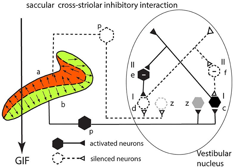
Have you ever wondered how you effortlessly maintain balance while navigating the world? It's not just magic—it's the intricate workings of your vestibular system, a sensory powerhouse hidden within your inner ear. One of the key players in this system is the saccule, a tiny organ with a big responsibility: sensing gravity and vertical movement. Let's take a closer look at how it accomplishes this remarkable feat, with a special focus on a fascinating process called cross-striolar inhibition, illustrated in the diagram below.
Reference: https://www.frontiersin.org/journals/neurology/articles/10.3389/fneur.2020.566895/full
The Saccule: Your Vertical Motion Sensor
Picture two tiny, fluid-filled pouches nestled deep within your inner ear—these are your saccules. Each saccule is a sensory marvel, lined with thousands of microscopic hair-like sensors (p) that dance and sway in response to gravity. These hair cells are arranged in two distinct sections:
Dorsal Section (a): The hair cells on this side are oriented to be inhibited (quieted down) when gravity (GIF) pulls downward.
Ventral Section (b): The hair cells on this side are oriented to be excited (activated) by the downward pull of gravity.
This clever arrangement allows the saccule to detect both the direction and magnitude of vertical movement.
Hair Cells and Neural Connections:
These hair cells are more than just sensors—they're connected to a network of neurons (black symbols) that transmit information to your brain.
Type I Hair Cells (p): These are the primary sensory cells, directly detecting movement and sending electrical signals to the brain via Type I neurons (c, d).
Type II Neurons (e, f): These are inhibitory neurons that modulate the activity of type I neurons, acting like traffic controllers to fine-tune the signals.
The Magic of Cross-Striolar Inhibition
Let's get to the heart of the matter: cross-striolar inhibition. When you tilt your head or move up and down, gravity (GIF) exerts a force on the saccule. This force causes the hair cells in the ventral section (b) to bend towards their tallest point, leading to their excitation. At the same time, the hair cells in the dorsal section (a) bend away from their tallest point, resulting in their inhibition.
Instead of these opposing signals canceling each other out, cross-striolar inhibition amplifies the difference between them. The excited hair cells (b) not only send strong signals to the brain via neuron (c) but also indirectly dampen the signals from the inhibited cells (a) via the inhibitory neuron (e). This creates a clearer, more robust signal that accurately reflects your body's movement.
A Walk in the Park, Revisited: Cross-Striolar Inhibition in Action
Imagine you're taking a leisurely stroll. With each step, gravity pulls down on your saccules. The hair cells on the bottom of the saccules get excited, while the ones on top get inhibited. Cross-striolar inhibition kicks in, boosting the signals from the excited cells and suppressing the signals from the inhibited cells. This tells your brain that your head is upright and your body is moving forward.
No Need for Cross-Talk: The Absence of Commissural Inhibition
Unlike the utricle, the saccule doesn't rely on communication with its counterpart on the other side of your head. This is because each saccule operates independently, expertly detecting vertical movement on its own.
Conclusion
Cross-striolar inhibition is a remarkable example of how our bodies have evolved ingenious ways to sense and interpret the world around us. It allows the saccule to provide our brains with precise information about our position and movement, helping us maintain balance, coordination, and spatial awareness. So the next time you step, jump in the air, or stand still, remember the incredible work your saccules are doing behind the scenes, keeping you grounded and balanced.
Reference and Further Reading
Curthoys IS. The Anatomical and Physiological Basis of Clinical Tests of Otolith Function. A Tribute to Yoshio Uchino. Front Neurol. 2020 Oct 20;11:566895. doi: 10.3389/fneur.2020.566895. PMID: 33193004; PMCID: PMC7606994.
https://www.frontiersin.org/journals/neurology/articles/10.3389/fneur.2020.566895/full
Uchino, Y., Sato, H., Sasaki, M., Imagawa, M., Ikegaya, K., & Isu, N. (1999). Sacculocollic reflex arcs in cats. I. Responses of vestibular neurons to electrical stimulation of the saccular macula. Journal of Neurophysiology, 82(3), 1421-1435. https://journals.physiology.org/doi/full/10.1152/jn.1997.77.6.3003