

The management of benign paroxysmal positional vertigo (BPPV) remains a cornerstone of vestibular practice. While clinicians resolve most cases with standard canalith repositioning maneuvers, such as the Epley or Semont, 12.5% of patients remain refractory. These patients often cycle through multiple clinics, experiencing persistent disability and a significant healthcare burden.
A recent study published in Frontiers in Neurology explores a transition toward personalized medicine for these difficult cases. Researchers propose that individual anatomical variations in the semicircular canals likely cause standard maneuver failure. By combining high-resolution MRI with Computational Fluid Dynamics (CFD), the study aims to develop a tailored approach to treat refractory posterior canal BPPV.
The Problem of Anatomical Variation
Standard repositioning maneuvers rely on the assumption that the semicircular canals sit at approximately 90 degrees relative to one another. However, human anatomy rarely fits a perfect geometric ideal. Individual differences in the spatial arrangement of the canals create a mismatch between the patient’s physical structure and the angles used by clinicians in traditional manual maneuvers.
When a clinician performs a standard Epley maneuver on a patient with atypical canal orientation, the otoconia may not reach the utricle. Instead, the particles might settle in a different part of the canal or fail to clear the common crus, leading to treatment failure.
Computational Fluid Dynamics and MRI Integration
The study employs a sophisticated workflow to bridge the gap between anatomy and successful repositioning.
3D Reconstruction from MRI
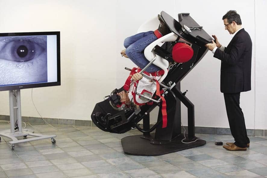
The process begins with a fluid-sensitive T2-weighted MRI. This scan includes the eyeballs to establish the ‘Frankfurt plane’ as a reference point. From these images, researchers reconstruct a 3D model of the endolymphatic labyrinth.
Fluid and Particle Simulation
The team applies CFD techniques to model the interaction between the endolymph and the otoconia. They calculate fluid velocity and pressure distribution by analyzing the density, viscosity, and gravity in the context of endolymph.
By coupling these calculations with a discrete particle model, the software predicts the exact trajectory of otoconia during a maneuver. This simulation accounts for gravity, fluid drag, and even the inertial forces generated by the patient’s body rotation.
The 100,000 Dollar Question: Chair vs Table
High-tech tools such as the Thomas Richard Vitton (TRV) chair or the Epley Omniax offer extreme precision, but their high cost makes them inaccessible to many private practices. The real value of this research lies in its application to the standard treatment table.
The study identifies that even a 9-degree variation in canal orientation can render a standard Epley maneuver ineffective. While a mechanical chair guarantees this precision, an expert clinician can manually replicate these specific angles. If we understand the specific angular deviations revealed by imaging, we can adapt our hands to match the math.
Key Components of Personalized Manual Maneuvers
If we apply this personalized medicine approach without a mechanical chair, we must focus on three specific areas:
Anatomical Mapping: We use MRI or high-resolution imaging to identify the patient’s specific canal plane relative to the ‘Frankfurt plane’.
Angular Modification: We adjust the degree of head rotation and extension during the Dix-Hallpike and Epley based on the individual’s unique geometry.
Inertial Control: We regulate the velocity of movement to ensure that the otoconia clear the common crus without falling back into the long arm of the canal.
Clinical Implications for Vestibular Professionals
This research moves BPPV treatment beyond the ‘one size fits all’ methodology. For the refractory patient, the issue may not be the diagnosis, but the geometry. Using CFD to predict otoconia movement enables clinicians to identify why a particle might become lodged and how to tilt the patient to dislodge it.
While CFD and MRI-based modeling currently require significant resources, they represent a frontier in personalized otoneurology. This approach could eventually reduce the need for invasive surgical options like canal plugging and provide a path to recovery for the most complex vestibular patients. We do not necessarily need a robot to cure refractory BPPV; we need better data on the patient’s internal structure.
The researchers conclude that by adjusting head movement angles to match specific anatomy, we can achieve faster symptom resolution and reduce the functional disability associated with persistent vertigo.
Reference
Rossi-Izquierdo M, Santos-Pérez S, Arán-Tapia I, et al. Personalized medicine to treat refractory benign paroxysmal positional vertigo, through computational fluid dynamics analysis from magnetic resonance image reconstructions. Front Neurol. 2025;16:1561356. Published 2025 Mar 11. doi:10.3389/fneur.2025.1561356