


Posterior Vitreous Detachment (PVD): A Vestibular Perspective

Mrs. Jones enters your clinic with a complaint of dizziness. 'Every time I lie down,' she explains, 'the room starts spinning.' You quickly recognize the hallmarks of Benign Paroxysmal Positional Vertigo (BPPV). But as you gather more information, a new layer of complexity emerges.
'My eye doctor told me I have something called... PVD?' she says, a hint of uncertainty in her voice. 'He said to be careful about lying flat.'
PVD? Posterior Vitreous Detachment. You mentally review your knowledge of eye conditions, but its relevance to vestibular treatment isn't immediately apparent. Mrs. Jones continues, 'I'm worried about those treatments where you have to lie back with your head over the edge of the table. Could that affect my PVD?'
This question, though common, often catches vestibular professionals off guard. As the population ages, we're encountering more patients with PVD, an eye condition that can mimic and complicate vestibular disorders. Understanding PVD is not just important but crucial for our practice.

What is PVD?
The vitreous humor is a clear, gel-like substance that fills the posterior cavity of the eye, between the lens and the retina. It's primarily composed of water, collagen, and hyaluronic acid. The vitreous plays a crucial role in:
Maintaining eye shape: Consider it the 'scaffolding' that helps the eye hold its form.
Supporting the retina: The vitreous gently presses the retina against the back of the eye, ensuring it stays in place.
Transmitting light: Its transparency allows light to pass through to the retina.
With age, the vitreous undergoes a process called syneresis. This means it gradually liquefies and shrinks, and the collagen fibers within it can clump together. As the vitreous shrinks, it can pull away from the retina, causing a posterior vitreous detachment (PVD).

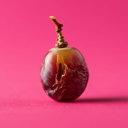
Think of it like this: Imagine a grape slowly drying out and shrinking. The skin of the grape (the vitreous) starts to separate from the inside (the retina). Sometimes, this separation happens cleanly. Other times, the skin can stick to the inside in certain spots, potentially causing a tear.
In the eye, this 'sticking' can create traction on the retina, leading to visual disturbances like floaters (the clumps of collagen casting shadows) and flashes of light (caused by the vitreous tugging on the retina). In some cases, the traction can be strong enough to cause a retinal tear or even a retinal detachment, a serious condition that can lead to vision loss.
Those Pesky Floaters
One of the most common symptoms of PVD is the appearance of floaters – those annoying little specks, strands, or cobweb-like shapes that drift across your field of vision. Floaters are tiny clumps of collagen fibers once part of the vitreous gel. When the vitreous detaches, these fibers can cast shadows on the retina, creating the illusion of floating objects. While usually harmless, a sudden increase in floaters, especially when accompanied by flashes of light, warrants immediate ophthalmological evaluation as it could signal a retinal tear.
In most cases, PVD itself does not require treatment.
Can PVD Be Treated?
In most cases, PVD itself does not require treatment. No medications, eye exercises, or dietary changes can reverse the process or eliminate floaters. However, it is crucial to monitor for potential complications, such as retinal tears. If a tear is detected, the ophthalmologist might recommend laser treatment or surgery to prevent it from progressing to a retinal detachment.
In rare cases where floaters significantly impair vision and become disabling, a surgical procedure called a vitrectomy (removal of the vitreous) might be considered. However, this is typically a last resort due to the potential risks associated with eye surgery.
Why Should Vestibular Professionals Care About PVD?
While PVD is primarily an ophthalmological condition, it can significantly impact the vestibular system. The intricate connection between vision and balance means that disturbances in one system can often manifest as symptoms in the other.
When the vitreous gel pulls away from the retina, it can stimulate sensory receptors, leading to symptoms overlapping with common vestibular disorders. These can include:
Dizziness: A general sense of unsteadiness or imbalance.
Vertigo: A distinct sensation of spinning or whirling, often triggered by head movements.
Visual disturbances: Floaters, flashes of light, and blurred vision can further contribute to disorientation and imbalance.
Differentiating PVD from Vestibular Disorders
So, how can we tell if a patient's dizziness is related to PVD or a primary vestibular disorder? Here are some key points to consider:
Thorough history: Ask about the onset and nature of their dizziness, any associated visual disturbances, and the specifics of their PVD diagnosis (including the presence of any retinal tears or restrictions from their ophthalmologist).
Oculomotor examination: Assess for any nystagmus (involuntary eye movements), which can indicate a vestibular problem. However, remember that some eye movements, like saccadic intrusions, can also be associated with PVD.
Positional testing: Carefully perform positional tests like the Dix-Hallpike maneuver, but modify them as needed to minimize stress on the retina. Observe if the dizziness is triggered explicitly by certain head positions, which might suggest BPPV.
Other vestibular tests: If safe and appropriate, consider tests like the Head Impulse Test to assess the vestibulo-ocular reflex (VOR).
Response to treatment: If the dizziness improves with vestibular rehabilitation exercises, it's less likely to be solely due to PVD.
Collaboration is Key
In many cases, it can be challenging to definitively determine whether dizziness is solely due to PVD, a primary vestibular disorder, or a combination of both. This is where your role as a vestibular professional becomes crucial. Open communication with the patient's ophthalmologist is important and integral to a comprehensive understanding of their eye health and any potential risks associated with vestibular assessment and treatment.
Assessment Challenges and Modifications
The Dix-Hallpike test, a key for BPPV, involves rapid head movements that could potentially worsen a retinal tear or even trigger a retinal detachment in someone with PVD. This underscores the need for caution and modification in vestibular tests for patients with PVD, promoting patient safety.
Furthermore, certain maneuvers, like the Semont, Gufoni, and Lempert Roll, could cause a transient rise in intraocular pressure (IOP). This is especially important in patients with glaucoma or other eye conditions sensitive to pressure changes.
Therefore, a thorough patient history is crucial. Ask about the specifics of their PVD diagnosis, any history of retinal tears, glaucoma, or other eye conditions, and any recommendations from their ophthalmologist.
If there's any concern, consider these modifications:
Modified Dix-Hallpike: To minimize pressure changes, perform the test with slower, more controlled movements.
Monitor for Valsalva: Watch for breath-holding or straining during the Dix-Hallpike, as this can increase IOP.
Alternative Assessments: In high-risk patients, favor assessments that don't involve head-down positioning.
Repositioning with Care
Even if BPPV is diagnosed, the standard Epley and Semont maneuver might need modification. Slower, gentler movements are key to ensuring the safety of the patient.
PVD: A Call for Interprofessional Collaboration
PVD adds another layer of complexity to vestibular assessment and management. By understanding the potential risks and modifying our approach, we can ensure the safety and well-being of our patients while providing effective vestibular rehabilitation. This highlights the importance of interprofessional collaboration between vestibular specialists and ophthalmologists to optimize patient care.
References
Bhattacharyya N, Gubbels SP, Schwartz SR, Edlow JA. Clinical Practice Guideline: Benign Paroxysmal Positional Vertigo (Update). Otolaryngol Head Neck Surg. 2017;156(3_suppl):S1-S47.
American Academy of Ophthalmology. Posterior Vitreous Detachment. [Internet]. San Francisco: American Academy of Ophthalmology; 2023 [cited 2025 Jan 17]. Available from: https://www.aao.org/eye-health/diseases/what-is-posterior-vitreous-detachment
Johnson MW. Posterior vitreous detachment: evolution and complications of its pathogenesis. Am J Ophthalmol. 1988;106(4):565-572.
Kanski JJ, Bowling B. Clinical Ophthalmology: A Systematic Approach. 9th ed. Edinburgh: Elsevier; 2017.
American Society of Retina Specialists. Posterior Vitreous Detachment. [Internet]. Chicago: American Society of Retina Specialists; 2023 [cited 2025 Jan 17]. Available from: https://www.asrs.org/patients/retinal-diseases/9/posterior-vitreous-detachment
Posterior Vitreous Detachment - Northside Eye Specialists. https://www.northsideeyespecialists.com/services/retinal-conditions/posterior-vitreous-detachment/