
Re-Engaging the Inner Ear: Precision Optokinetics for Unilateral Vestibular Hypofunction – Part 1: The ‘Torque’ of Adaptation


Here is the scenario: You have done everything ‘by the book.’ You work diligently with a unilateral vestibular hypofunction (UVH) patient and perform gaze stabilization exercises. You progress from VOR x1 to VOR x2, even incorporating unilateral head impulse training (UHIT). Yet, despite your best efforts, something feels stuck. You put on those infrared (IR) goggles (VestibularFirst/Vesticam) and see it: persistent spontaneous nystagmus, a tell-tale sign that your patient has not fully achieved static compensation, let alone dynamic.

The clinical plateau frustrates you, and you search for that next powerful tool to drive progress as your patient doesn’t feel right - they feel off, frustrated as well.
What if you could induce a profound, corrective ‘torque’ on the vestibular system, not by spinning the patient or doing the VOR x1/x2 but by masterfully controlling their visual environment?
Enter optokinetic stimulation (OKS). This is not just a visual exercise but a potent driver of vestibular adaptation through vection – the compelling illusion of self-motion a moving visual field induces. As physical therapists specializing in balance and dizziness disorders, we often access systems capable of generating robust optokinetic flows through projected Bárány bars or other digital displays. Still, the precise application remains a mystery for many.
In this two-part series, we will demystify this powerful approach. This part (Part 1) will lay the foundational understanding: the neurophysiology of vestibular hypofunction, how OKS generates a profound ‘torque’ on the vestibular system via vection, and why this is a critical mechanism for inducing adaptation in patients with UVH. This concept is not dissimilar to other forms of forced vestibular stimulation, such as caloric irrigation or rotational chair testing, but it offers unique advantages in a clinical setting.
The Asymmetrical Challenge: Understanding Unilateral Vestibular Hypofunction
Our balance system relies on symmetrical input from both inner ears. A healthy individual’s tonic (resting) firing rate from the vestibular afferents on both sides is equal. When the head moves, one side’s firing rate increases while the other’s decreases, providing the brain with a precise ‘push-pull’ signal of head velocity.
In unilateral vestibular hypofunction (UVH), this delicate symmetry is disrupted. Due to injury, disease, or aging, one inner ear’s function diminishes. This leads to a constant, uncompensated asymmetry in the tonic firing rates from the vestibular nuclei in the brainstem. The brain perceives a ‘drift’ or ‘spin’ that is not occurring, leading to debilitating symptoms:
Vertigo: A false sensation of spinning, tilting, or rocking.
Oscillopsia: The visual world appears to jiggle or blur, especially during head movement, due to an impaired vestibulo-ocular reflex (VOR).
Postural Instability: Patients experience difficulty maintaining balance, particularly in challenging vision and surface environments.
The central nervous system, always striving for symmetry, attempts to compensate. However, this process often needs a precise, directed therapeutic push.
The ‘Torque’ Concept: How Optokinetics Drives Adaptation through Vection
Imagine the vestibular system as a finely balanced gyroscope. When one side is underactive, the gyroscope is off-kilter. Our goal in rehabilitation is to re-level it, or more accurately, to teach the brain to operate effectively with the new, reduced input. We achieve this by creating a compensatory ‘torque’ – a force that encourages the brain to re-establish a new, functional equilibrium.
How do we produce ‘torque’ on the vestibular system and VOR? We induce this ‘torque’ through direct vestibular stimulation:

Forced Spinning: Rotational chair (computer chair) or therapeutic maneuvers (like the Epley or Li maneuver for BPPV) generate direct angular acceleration. We use short, intense spins for specific canal stimulation and longer, sustained spins to drive broader adaptive changes. These spins stimulate the semicircular canals and produce a powerful vestibular signal.

Caloric Stimulation: This method applies thermal stimulation (cold or warm air/water) to the ear canal. The temperature difference creates convection currents within the endolymph of the horizontal semicircular canal, mimicking head movement and inhibiting (cold) or exciting (warm) the vestibular system. This induces an intense ‘spin’ sensation.

Head Shaking Test: Rapid, repeated head movements create sustained angular acceleration, challenging the vestibular system.

Unilateral Hepulse Training: Predictable, high-velocity head thrusts specifically towards the affected ear side directly engage the VOR, forcing it to work rapidly and adapt.
These methods directly ‘force’ a vestibular response.
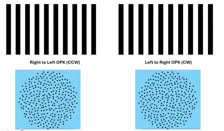
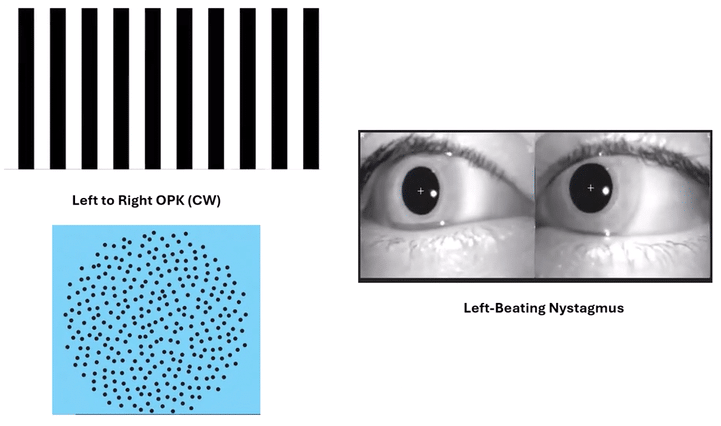
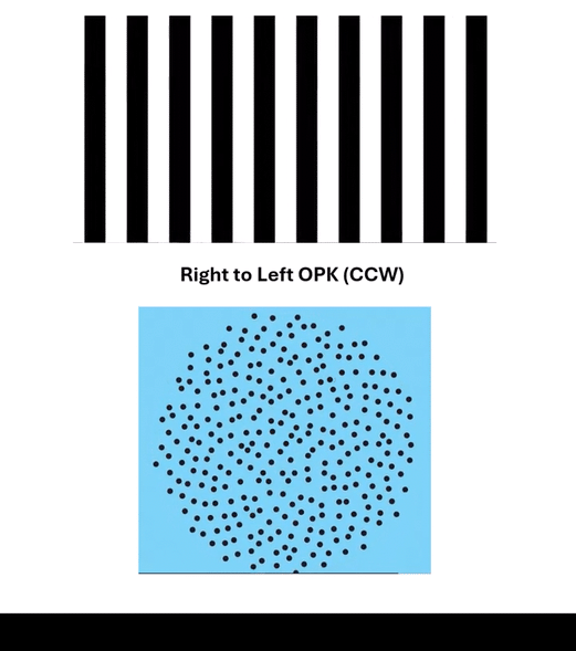
Now, consider optokinetic stimulation (OKS). While it begins as a visual stimulus, its far-reaching effect extends deeply into the vestibular system. When a person observes a moving full-field visual environment (like moving stripes), their eyes automatically perform optokinetic nystagmus (OKN) – a reflexive eye movement consisting of a slow phase that follows the visual stimulus and a rapid, corrective fast phase in the opposite direction.
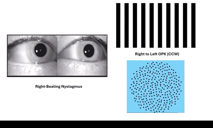
Here is the critical connection: this visually driven OKN is not just about the eyeballs. The continuous slow-phase movement of the eyes, driven by the optokinetic stimulus, feeds into the same neural pathways that process vestibular information. The brain interprets this sustained visual ‘flow’ as if the body is rotating. This sustained visual input creates a powerful, corrective ‘torque’ on the central vestibular system, effectively mimicking and driving the exact adaptive mechanisms that direct vestibular stimulation would. This compelling illusion of self-motion is known as vection.
This ‘torque’ via vection impacts the entire vestibular network, influencing reflexes at multiple levels:
Vestibulo-Ocular Reflex (VOR): This is the most immediate and noticeable effect. OKS trains and recalibrates the VOR, crucial for gaze stability during head movements. By continually forcing eye movements in a specific direction (fast phase towards the paretic side in UVH), we actively promote gain adaptation of the VOR on that side.
Vestibulocollic Reflex (VCR): This reflex acts on the neck muscles to stabilize the head in space. The ‘torque’ OKS extends to the VCR, helping to re-establish proper head stabilization responses relative to the body, even when direct vestibular input diminishes.
Vestibulospinal Reflex (VSR): Crucially, the ‘torque’ extends to the body’s postural muscles through the VSR. This reflex helps maintain balance and posture. By driving this visually-induced ‘self-motion’ signal through vection, OKS helps to re-weight and recalibrate the postural control system, improving standing and walking balance.
The Interplay of Reflexes: Forcing Deeper Adaptation
We can further amplify the adaptive power of OKS by strategically challenging the lower-level vestibular reflexes. The balance system operates hierarchically. When we diminish the effectiveness of the Vestibulospinal Reflex (VSR) – the reflex most directly responsible for maintaining gross body posture – we force the higher-level reflexes, the VCR and VOR, to work harder.
For instance, when a patient performs an activity on a compliant or uneven surface or stands on a narrow base of support, we directly challenge the VSR and the somatosensory system. This actively ‘disengages’ or makes the most common postural compensatory strategies less reliable. This forces the brain to place greater reliance and demand on the VCR and VOR to maintain stability and accurate gaze. By combining this postural challenge with the direct ‘torque’ of OKS, we create a powerful synergistic effect. This pushes the entire vestibular network to adapt more effectively and solidify the desired neuroplastic changes, transforming static compensation into robust dynamic function.
The optokinetic flow produces a sustained, directional bias in the brainstem vestibular nuclei. Moving stripes from right to left generates a consistent ‘pull’ or ‘torque’ to the right for a patient with a right unilateral hypofunction. This continuous, forced activation helps ‘reset’ the pathological asymmetry, promoting neuroplastic changes that improve compensation. It is an active, powerful stimulus that directly engages the brain’s adaptive machinery, like how a foam cushion directly challenges the somatosensory system to encourage adaptation.
Looking Ahead to Part 2
Understanding the ‘why and what’ behind OKS for UVH is foundational. In Part 2 of this series, we will delve into the practical ‘how’ – the specific parameters we manipulate, such as velocity, bar width, exposure time, and environmental factors like surface and base of support, to precisely dose and optimize this powerful adaptive tool in our clinical practice. This will provide you with actionable strategies to implement precision optokinetics effectively.