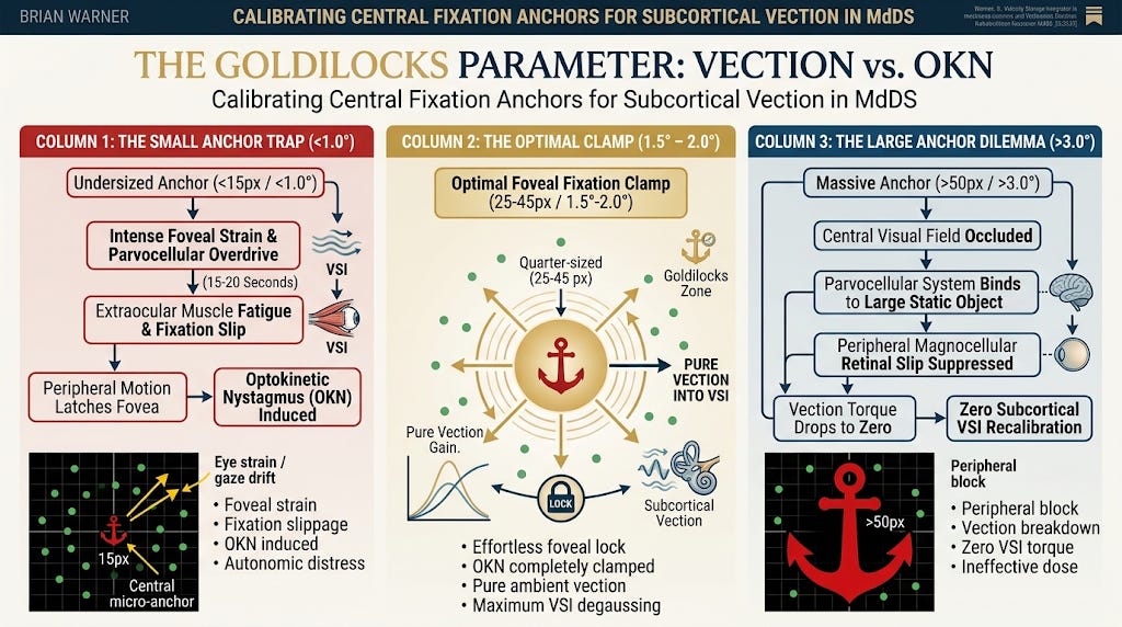
The Goldilocks Parameter: Calibrating Central Fixation Anchors for Subcortical Vection in Mal de Debarquement Syndrome (MdDS)

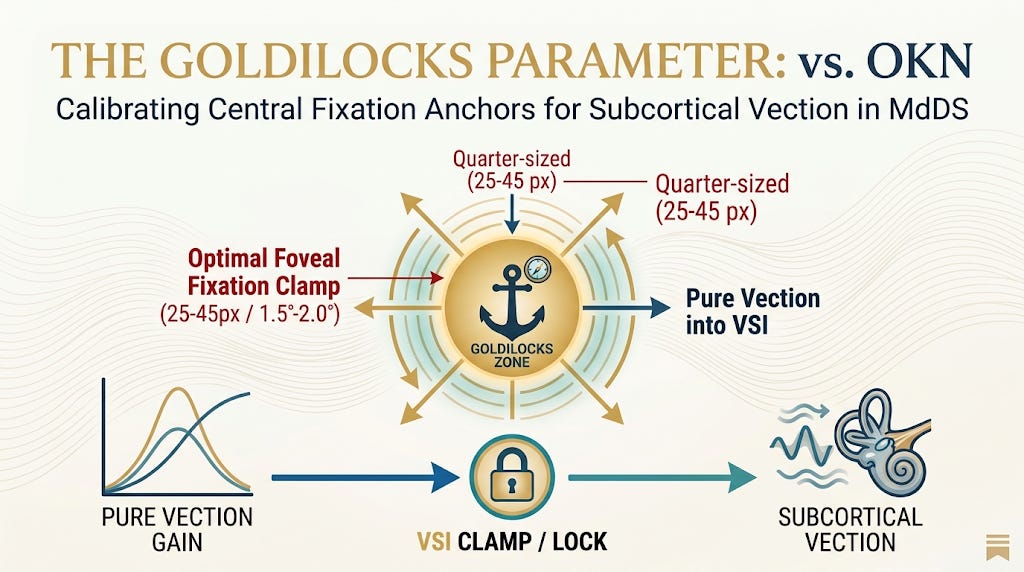
In the non-pharmacological rehabilitation of Mal de Débarquement Syndrome (MdDS), leveraging optokinetic flow to recalibrate the Velocity Storage Integrator (VSI) represents a major paradigm shift away from simple habituation or adaptation (Dai et al., 2014, 2017). However, clinical success hinges upon a precise biophysical trade-off: foveal fixation versus peripheral motion perception (Frattini & Wibble, 2021; Pola et al., 1995).
To deliver therapeutic visual torque without triggering acute autonomic distress or visual motion sickness, clinicians must control the spatial geometry of the central fixation target—the Visual Anchor (Nooij et al., 2018).
Modulating the physical scale of this anchor dictates whether the central nervous system successfully engages in subcortical degaussing or collapses into ocular strain and therapy breakdown.
1. The Small Anchor Trap: Oculomotor Fatigue and OKN Induction
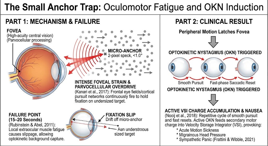
When the visual anchor is calibrated below functional thresholds (e.g., a tiny 2-pixel speck or an unsharpened micro-point), the fovea—the central visual field responsible for high-acuity, parvocellular processing—must exert significant effort to sustain fixation (Pola et al., 1995; Rubinstein & Abel, 2011).
The Mechanism:
To prevent the eyes from naturally locking onto the surrounding moving dot field, the frontal eye fields and cortical pursuit networks must continuously fire to hold the fovea on an undersized target (Kanari et al., 2017).
The Failure Point:
Within 15 to 20 seconds, local extraocular muscle fatigue causes fixation “slippage.” The gaze drifts off the micro-anchor, allowing the surrounding optokinetic background to capture central vision (Rubinstein & Abel, 2011).
The Clinical Result:
Foveal capture triggers Optokinetic Nystagmus (OKN)—a repetitive cycle of smooth pursuit and fast-phase saccadic resets (Nooij et al., 2018). Instead of clearing the trapped subcortical spatial error, active OKN feeds a secondary motor charge into the velocity storage integrator, potentially provoking acute motion sickness, migrainous head pressure, and sympathetic panic (Frattini & Wibble, 2021; Nooij et al., 2018).
2. The Large Anchor Dilemma: Foveal Occlusion and Vection Decay
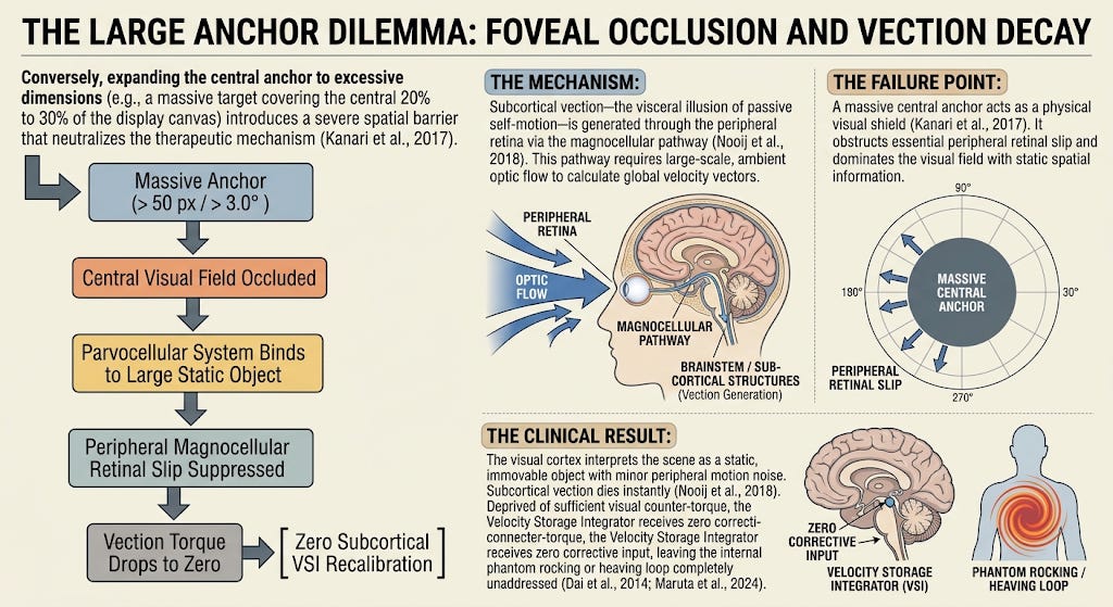
Conversely, expanding the central anchor to excessive dimensions (e.g., a massive target covering the central 20% to 30% of the display canvas) introduces a severe spatial barrier that neutralizes the therapeutic mechanism (Kanari et al., 2017).
The Mechanism:
Subcortical vection—the visceral illusion of passive self-motion—is generated through the peripheral retina via the magnocellular pathway (Nooij et al., 2018). This pathway requires large-scale, ambient optic flow to calculate global velocity vectors.
The Failure Point:
A massive central anchor acts as a physical visual shield (Kanari et al., 2017). It obstructs essential peripheral retinal slip and dominates the visual field with static spatial information.
The Clinical Result:
The visual cortex interprets the scene as a static, immovable object with minor peripheral motion noise. Subcortical vection dies instantly (Nooij et al., 2018). Deprived of sufficient visual counter-torque, the Velocity Storage Integrator receives zero corrective input, leaving the internal phantom rocking or heaving loop completely unaddressed (Dai et al., 2014; Maruta et al., 2024).
3. The Goldilocks Parameter: Achieving the Foveal Clamp
To optimize therapeutic outcome, the visual anchor must be set to a precise subcortical “sweet spot”—a configuration known as the Foveal Fixation Clamp (Pola et al., 1995).
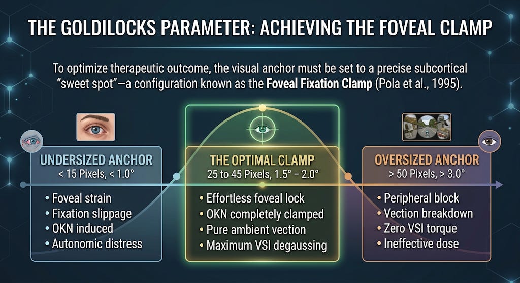
Clinical Calibration Standards:
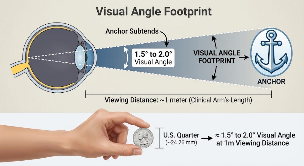
Visual Angle Footprint:
The anchor must subtend 1.5° to 2.0° of visual angle. At standard clinical arm’s-length viewing distance (~1 meter), this translates to an object approximately the size of a quarter.
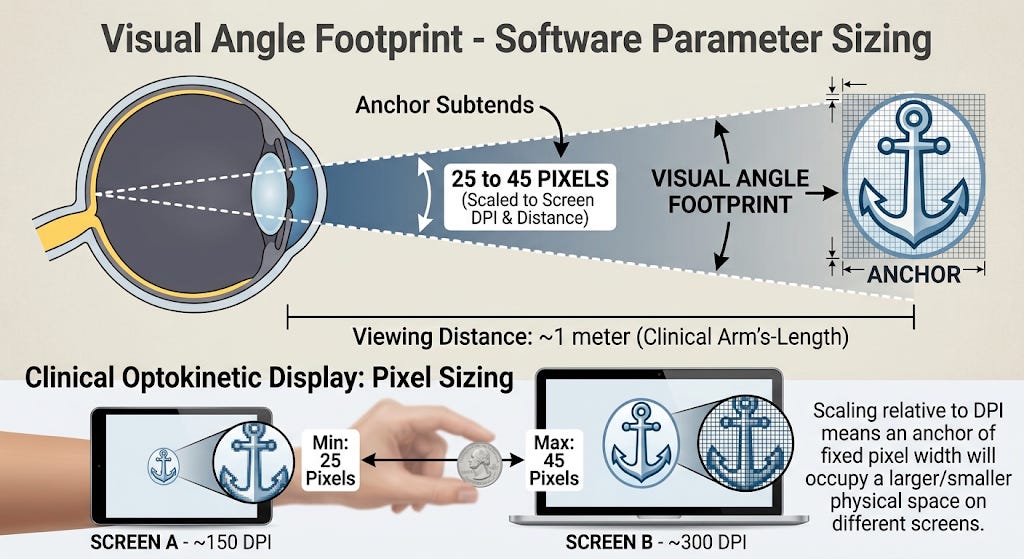
Software Parameter Sizing:
In high-resolution digital optokinetic displays, this corresponds to an Anchor Size of 25 to 45 pixels (scaled relative to screen DPI and viewing distance).
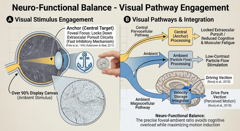
Neuro-Functional Balance:
This specific scale provides enough foveal weight to lock down extraocular pursuit circuits using fast inhibitory mechanisms without causing cognitive or muscular fatigue (Pola et al., 1995; Rubinstein & Abel, 2011). Simultaneously, it leaves over 90% of the display canvas open for low-contrast particle flow to stimulate the ambient magnocellular pathway and drive pure vection into the Velocity Storage Integrator (Nooij et al., 2018).
Audited Reference List
Dai, M., Cohen, B., Cho, C., Shin, S., & Yakushin, S. B. (2014). Readaptation of the vestibulo-ocular reflex relieves the mal de debarquement syndrome. Frontiers in Neurology, 5, Article 124.
Dai, M., Cohen, B., Cho, C., Shin, S., & Yakushin, S. B. (2017). Treatment of the Mal de Debarquement Syndrome: A 1-year follow-up. Frontiers in Neurology, 8, Article 175.
Frattini, D., & Wibble, T. (2021). Alertness and visual attention impact different aspects of the optokinetic reflex. Investigative Ophthalmology & Visual Science, 62(13), Article 16.
Kanari, K., Sakamoto, K., & Kaneko, H. (2017). Effect of visual attention on the properties of optokinetic nystagmus. PLOS ONE, 12(4), Article e0175453.
Maruta, J., Cho, C., Raphan, T., & Yakushin, S. B. (2024). Symptom reduction in mal de débarquement syndrome with attenuation of the velocity storage contribution in the central vestibular pathways. Frontiers in Rehabilitation Sciences, 5, Article 1331135.
Nooij, S. A. E., Pretto, P., & Bülthoff, H. H. (2018). More vection means more velocity storage activity: A factor in visually induced motion sickness? Experimental Brain Research, 236(11), 3031–3041.
Pola, J., Wyatt, H. J., & Lustgarten, M. (1995). Visual fixation of a target and suppression of optokinetic nystagmus: Effects of varying target feedback. Vision Research, 35(8), 1079–1087.
Rubinstein, N. J., & Abel, L. A. (2011). Optokinetic nystagmus suppression as an index of the allocation of visual attention. Investigative Ophthalmology & Visual Science, 52(1), 462–467.