

The Runaway Flywheel Versus the Hyper-Vigilant System: Decoding the Architecture of Resetting MdDS and PPPD


The clinical frontier of central vestibular disorders requires a profound shift away from classic peripheral rehabilitation models. When managing complex presentations like Mal de Débarquement Syndrome (MdDS) and Persistent Postural-Perceptual Dizziness (PPPD), clinicians are not treating a structural deficit of the inner ear. Instead, they are manipulating a central processing error. While both populations present with severe spatial disorientation, chronic sensory mismatch, and debilitating autonomic distress, their underlying neuroanatomical architecture is diametrically opposed.
To address these conditions, clinicians must employ highly specific subcortical protocols.
By understanding how these networks interact with central gamma-aminobutyric acid (GABA) receptors, we can execute targeted clinical interventions through The Zero Øut Method™ to systematically reset both systems.
The Neurological Divergence: Locked Flywheel vs. Clamped Flywheel
To understand the treatment solutions for these disorders, we must first isolate the distinct behavioral states of their central integrators.
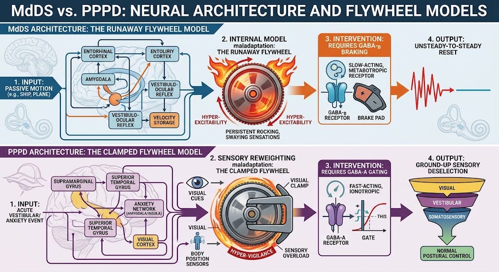
In MdDS, the brainstem’s velocity storage integrator—the neural flywheel that naturally prolongs our central perception of low-frequency motion—is running out of control.
Trapped in an autonomous, continuous, low-frequency oscillation (typically 0.2 to 0.3 Hz), this flywheel perpetually discharges an internal motion error up the vestibulothalamocortical pathway. The system is locked in a self-sustaining loop independent of actual peripheral input. This persistent error triggers chronic hypermetabolism in the left entorhinal cortex and drives the amygdala into an intense state of survival-based hypervigilance.
In PPPD, the underlying architecture is entirely different.
The velocity storage integrator and its neural flywheel are structurally and functionally intact. However, the central nervous system has sustained a profound loss of confidence in its internal inertial models, frequently secondary to an acute vestibular event or a period of severe physiological threat.
To protect the organism, the brainstem and cortex clamp down on the velocity storage network, refusing to access the automatic flywheel.
Instead of relying on subconscious equilibrium, the central nervous system switches to a highly rigid, voluntary postural control strategy. The system operates with exaggerated visual and somatosensory components, overanalyzing every micron of postural sway. The system interprets normal environmental variance as an immediate threat, completely overwhelming the fast-acting, ionotropic GABA-A gating networks that normally filter out background sensory noise.
The Kinetics of Inhibition: GABA-B vs. GABA-A
The distinct receptor kinetics of the central nervous system dictate how these two pathways must be downregulated.
The velocity storage network is modulated primarily by metabotropic, G-protein-coupled GABA-B receptors located along the crossing commissural pathways of the brainstem vestibular nuclei.
GABA-B receptors exhibit slow, sustained hyperpolarization kinetics, making them uniquely suited to serve as the primary physiological brake on a low-frequency, long-duration system such as the neural flywheel.
In MdDS, driving a massive influx of visual motion data through the magnocellular pathway forces these commissural fibers to release a profound wave of GABA-B inhibition, effectively putting a physical stop to the flywheel’s runaway momentum.
Conversely, the fast sensory gating and immediate filtering of background environmental noise rely on ionotropic GABA-A receptors.
These receptors open chloride channels almost instantly, providing rapid, stimulus-driven, short-duration inhibition. In the hyper-vigilant state of PPPD, these fast-acting GABA-A gating networks are completely overwhelmed by the brain’s frantic attempt to voluntarily process every piece of incoming visual and somatosensory data. To quiet PPPD, the clinician must implement protocols that reduce the computational load on these fast channels, signal safety to the GABA-A infrastructure, and allow the cortex to release its rigid, voluntary grip on the brainstem.
The Treatment Solution for MdDS: The Top-Down, Unsteady-to-Steady Deceleration
Because the MdDS flywheel is spinning out of control, the treatment strategy requires a powerful, top-down subcortical override paired with an Unsteady-to-Steady postural deceleration mechanism.
The clinical objective is to capture the runaway integrator using high-demand optokinetic flow to produce vection, while systematically driving the client from a state of high physical unsteadiness down to a state of max somatosensory stability.
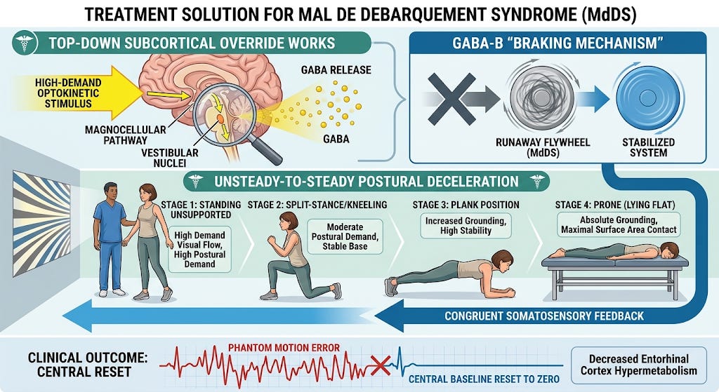
The execution begins with the client standing without support, directly exposed to a tailored optokinetic stimulus designed to match and counter the internal frequency of their central oscillation. This high-demand visual motion aggressively fires the magnocellular pathway, delivering massive optical flow data to the vestibular nuclei. As the magnocellular input creates a controlled vection torque, it stimulates the cross-brainstem commissural neurons to release a profound wave of GABA, engaging the slow-acting GABA-B receptors to apply the brakes to the runaway flywheel.
Simultaneously, the clinician executes the postural reset by taking the client through a progressive reverse mechanism. The client moves systematically from standing without support to a split-stance or kneeling position, then to a highly stable plank position, and ultimately to lying prone on their stomach, flat on the surface.
By forcing the body from a state of high postural demand to an absolute, high-surface-area grounding, the clinician delivers an undeniable wave of congruent somatosensory feedback.
This rapid transition from unsteady to steady reinforces the subcortical GABA-B braking mechanism, completely crushing the phantom 0.2 Hz motion error, lowering the hypermetabolism in the left entorhinal cortex, and resetting the central baseline back to zero.
The Treatment Solution for PPPD: The Ground-Up Sensory Deselection Protocol
Quieting the hyper-vigilant system of a client with PPPD requires a fundamentally different clinical trajectory. The objective is not to break a broken flywheel, but to systematically train the brain to ignore its exaggerated visual component through sensory substitution and gradual somatosensory deselection. This protocol moves from the ground up, moving from absolute zero back out into space.
The critical realization in this method is that both MdDS and PPPD populations require optokinetics and vection, but they utilize them for entirely different neurological purposes.
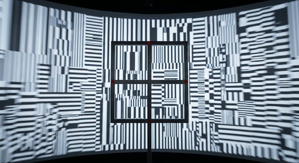
For the PPPD client, high-demand optokinetic flow is not used to brake an oscillator, but to deliberately introduce a visual torque to the balance system while the client is in a position of maximum mechanical advantage.
The protocol is executed by placing the client in a completely stable, supported position—initially lying flat on their back (supine) or side-lying on their right or left side. While flat against the surface, a large-field optokinetic stimulus is introduced. Crucially, the visual stimulus is designed with a specific architecture: a moving, dynamic background that induces vection, juxtaposed with a rigid, stationary foreground anchor that the client is instructed to stabilize visually.
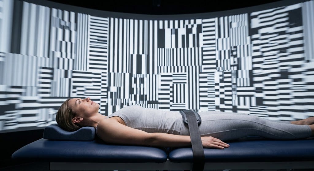
When this visual torque is introduced while the client is completely flat, it generates a deliberate sensory conflict. Because the client is lying down, their remaining somatosensory receptors and otolith organs provide an absolute, unchanging baseline of gravity and surface contact. This massive, grounded sensory inflow provides the central nervous system with enough definitive data to successfully override the visual illusion of movement.
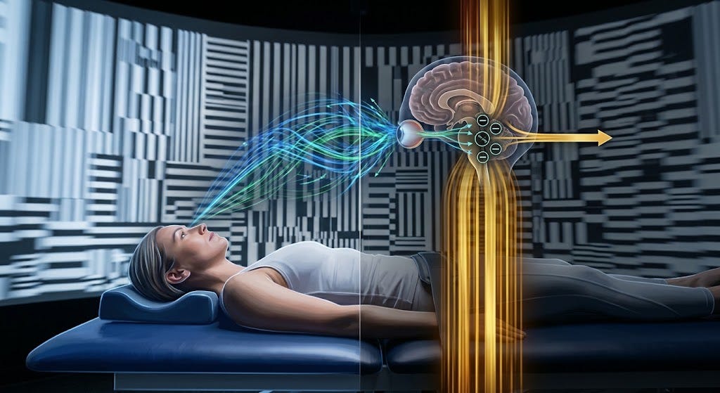
Rather than becoming desensitized to the visual flow through simple exposure, the brain actively employs a strategy of sensory substitution. It recognizes that visual motion is irrelevant to actual physical orientation, thereby using fast GABA-A gating networks to filter out background visual noise.
The client learns to entirely ignore the provocative visual input, downregulating the brain’s maladaptive visual dependency without triggering symptoms.
Once the client demonstrates the ability to maintain complete neural quietude and sensory substitution in the flat position, the clinician begins the systematic process of somatosensory deselection. The clinician slowly and progressively takes away the supportive somatosensory surface area, forcing the central nervous system to maintain its automatic gating control under higher postural demands:
Move the client from supine or side-lying to a prone position, flat on the stomach.
Progress to a prone-on-elbows position, elevating the neuraxis while maintaining forearm contact.
Advance to a full plank position, and subsequently execute side plank positions to reduce the lateral base of support.
Transition the client onto dynamic surfaces, such as lying supine on a Swiss ball, moving to prone bridging over a Swiss ball, and executing targeted bridging sequences.
By slowly disengaging the supportive somatosensory system while maintaining the optokinetic visual torque, the brain is forced to transfer its automatic gating strategies up the chain.
Because the client has already learned to ignore the visual conflict at baseline “zero,” the progressive reduction of surface contact safely coaxes the central nervous system to reopen and trust its normal velocity storage network.
The voluntary, rigid postural clamp is systematically dismantled, the hyper-vigilant amygdala stands down, and the client successfully returns to effortless, automatic, and subconscious equilibrium.
Verifiable Clinical References
Cha, Y. H. (2021). Neuroimaging markers of Mal de Débarquement Syndrome. Frontiers in Neurology, 12, Article 636224. https://doi.org/10.3389/fneur.2021.636224
Cha, Y. H., Chakrapani, S., Craig, A., & Baloh, R. W. (2012). Metabolic and functional connectivity changes in Mal de Debarquement Syndrome. PLoS ONE, 7(11), Article e49560. https://doi.org/10.1371/journal.pone.0049560
Dai, M., Cohen, B., Smouha, E., & Cho, C. (2014). Readaptation of the vestibulo-ocular reflex to modify Mal de Debarquement Syndrome. Frontiers in Neurology, 5, Article 124. https://doi.org/10.3389/fneur.2014.00124
Holstein, G. R., Martinelli, G. P., Wearne, S., & Cohen, B. (1999). The ultrastructure of GABA-immunoreactive vestibular commissural neurons related to velocity storage in the monkey. Neuroscience, 93(1), 171-181. https://doi.org/10.1016/s0306-4522(99)00147-1
Popa, L. S., Hewitt, A. L., & Ebner, T. J. (2014). Purkinje cell types and their alterations in vestibular disorders. Journal of Neurophysiology, 112(11), 2743-2753. https://doi.org/10.1152/jn.00421.2014
Yakushin, S. B., Xiang, Y., Holstein, G. R., & Cohen, B. (2017). Coding of velocity storage in the vestibular nuclei. Frontiers in Neurology, 8, Article 386. https://doi.org/10.3389/fneur.2017.00386
Professional Notice and Medical Disclaimer
The theoretical framework presented in this article regarding the specific subcortical interactions of the magnocellular pathway, GABA-B mediated commissural inhibition, and downstream cortical/limbic downregulation represents the clinical hypothesis and working model of Brian Keith Werner, MPT. While this framework systematically integrates established, peer-reviewed neuroanatomical data and functional neuroimaging findings from the cited literature, the unified synthesis and its direct application within The Zero Øut Method™ are proprietary theoretical concepts developed to better understand, model, and manage this challenging client population.
This content is published exclusively for educational, informational, and professional discussion purposes among vestibular specialists and healthcare providers. It does not constitute formal medical advice, diagnosis, or a guarantee of clinical outcomes. Vestibular professionals must use independent clinical judgment when evaluating and managing individuals experiencing signs or symptoms consistent with Mal de Débarquement Syndrome or Persistent Postural-Perceptual Dizziness.