


Introduction
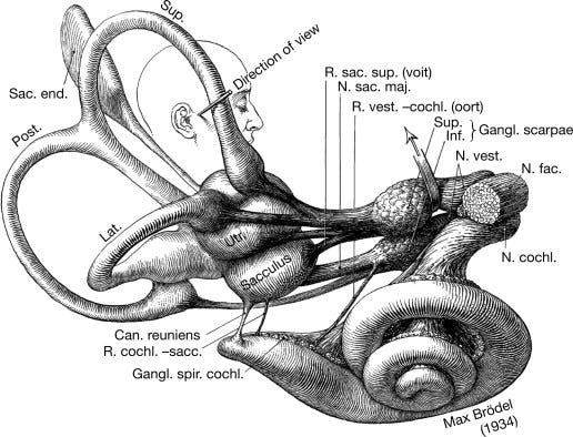
The semicircular canals are integral components of the vestibular system, responsible for our sense of balance, spatial orientation, and coordination of eye movements with head movements. These three tiny, fluid-filled tubes, nestled within the inner ear, play a crucial role in our daily lives, enabling us to walk, run, and interact with our environment seamlessly. Each canal is located in a different plane in space, with the anterior canal oriented vertically, the posterior canal oriented horizontally, and the lateral canal oriented at a 45-degree angle. This arrangement allows the canals to detect head movements in all three dimensions.
Anatomy of the Semicircular Canals
The semicircular canals are named according to their orientation:
Anterior (Superior) Canal: Detects head movement in the sagittal plane (nodding "yes").
Posterior Canal: Detects head movement in the coronal plane (tilting the head towards the shoulder).
Lateral (Horizontal) Canal: Detects head movement in the transverse plane (shaking "no").
Each canal has a dilated end called the ampulla, which houses the sensory epithelium known as the crista ampullaris. The crista ampullaris contains hair cells embedded in a gelatinous structure called the cupula. When the head moves, the endolymph in the canals lags behind due to inertia, causing the cupula to bend. This bending of the cupula stimulates the hair cells, sending signals to the brain that allow it to detect and interpret head movements.
Physiology of the Semicircular Canals
Head Movement: When the head rotates, the endolymph (fluid filling the canals) lags due to inertia. This lag in the endolymph allows the canals to detect head movements. The movement of the endolymph in response to head movements is a crucial part of the process by which the canals help us maintain our balance.
Endolymph Flow: Relative motion between the canals and the endolymph causes the fluid to flow, deflecting the cupula in the direction opposite to head movement.
Hair Cell Activation: The cupula deflection bends the hair cells' stereocilia (hair-like projections). When stereocilia bend towards the tallest stereocilium (kinocilium), it opens mechanically-gated ion channels, allowing positively charged ions (primarily potassium and calcium) to enter the cell.
Mechanoelectrical Transduction: This is where the real magic happens. The influx of ions depolarizes the hair cell, generating a receptor potential. The magnitude of this potential is proportional to the degree of hair cell deflection, which in turn corresponds to the velocity of head rotation. It's a complex process, but understanding it is critical to understanding the semicircular canals' function.
Neurotransmitter Release: The receptor potential triggers the release of neurotransmitters (primarily glutamate) at the base of the hair cell, where it forms synapses with the dendrites of vestibular nerve fibers.
Nerve Impulse Transmission: Neurotransmitters excite vestibular nerve fibers, initiating action potentials that propagate along the nerve to the brainstem.
Push-Pull Mechanism
The semicircular canals operate in a push-pull fashion. When the head rotates in one direction, the hair cells in the canal on that side are excited, while the hair cells in the corresponding canal on the opposite side are inhibited. This differential activation provides the brain with information about the direction and magnitude of head rotation. For example, when you turn your head to the right, the hair cells in the correct horizontal canal are excited, while the hair cells in the left are inhibited. This allows the brain to accurately perceive the direction of your head movement.
Central Processing of Vestibular Information
The vestibular nerve fibers project to the vestibular nuclei in the brainstem. These nuclei process the information and relay it to various brain regions, including the:
Cerebellum: Coordinates balance and movement.
Oculomotor Nuclei: Control eye movements (via the vestibulo-ocular reflex, VOR).
Spinal Cord: Modulates muscle tone and posture (via the vestibulospinal reflexes, VSR).
Thalamus: Relays vestibular information to the cerebral cortex for conscious perception of movement and spatial orientation.
Clinical Significance
Dysfunction of the semicircular canals can lead to various disorders, including:
Benign Paroxysmal Positional Vertigo (BPPV): Characterized by brief episodes of vertigo triggered by specific head positions caused by displaced otoconia in the semicircular canals.
Vestibular Neuritis: An inflammation of the vestibular nerve, causing dizziness, vertigo, and nausea.
Labyrinthitis: An infection of the inner ear, leading to hearing loss, vertigo, and tinnitus.
Meniere's Disease: A chronic inner ear disorder characterized by episodic vertigo, tinnitus, and hearing loss.
Diagnosis and Treatment
Vestibular disorders are typically diagnosed through a combination of:
Patient history and physical examination
Vestibular function tests (e.g., VEMPs, rotational chair testing, caloric testing)
Imaging studies (e.g., MRI)
Treatment options depend on the specific disorder, but may include:
Vestibular rehabilitation therapy
Medications (e.g., antiemetics, antihistamines)
Surgery (in rare cases)
Conclusion
The semicircular canals, these marvelously intricate structures, are fascinating to study and of utmost clinical significance. Understanding their anatomy and physiology is crucial for diagnosing and treating various vestibular disorders, which can significantly impact a person's quality of life. This underscores the importance of our study and research in this field.