
Updated: The Zero Out Method for Mal de Débarquement Syndrome: Overwriting the Corrupted Vestibular Software Patch


Mal de Débarquement Syndrome (MdDS) represents a severe, persistent failure of the central nervous system to readapt to a stationary land environment following prolonged exposure to passive transit motion (Brown & Baloh, 1987; Cha, 2009). The Zero Out Method provides a specialized neurotological rehabilitation framework engineered to systematically isolate, confront, and purge these deeply entrained, multi-axial motion patterns.

By employing a foveal anchor against structured peripheral visual stimulation, this methodology forces a precise top-down sensory mismatch. Rather than relying on slow, volatile habituation strategies, the protocol drives visual vection across sixteen distinct eigenvector directions while sequentially grounding the patient through the complete developmental postural hierarchy, using primitive somatosensory-vestibular networks to execute an absolute system reset.
The Low-Frequency Brainstem Filter Failure and Transit Soporific Entrainment
To understand why this corrupted neural loop becomes so deeply entrenched, the framework must isolate the brainstem's and vestibulocerebellum's specific low-frequency processing limits.

Commercial aircraft, passenger trains, and ocean vessels generate continuous, low-frequency passive oscillations that settle precisely between 0.2 Hz and 0.3 Hz. As established by Cohen, Yakushin, and Cho (2018), the human central nervous system processes these specific low frequencies through a specialized brainstem-cerebellar filter pathway localized to the nodulus and uvula. This network functions as a low-pass filter, dampening low-frequency otolithic translation data to protect the cerebral cortex from sensory saturation during prolonged environmental motion.
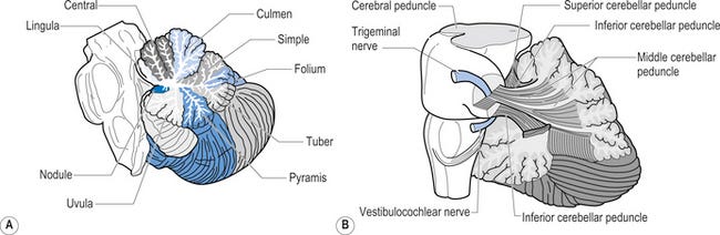
CLINICAL INSIGHT: The Brainstem Filter as a Permanent Switch. The nodulus and uvula act as the primary gatekeepers. When this filter over-adapts during transit, it does not just dampen signals—it locks the velocity storage integrator into an internal, 0.2–0.3 Hz loop that continues firing long after the physical stimulus has ceased.
This low-frequency stimulation drives entrainment whether the passenger is wide awake or asleep. When a passenger is awake, their foveal vision is locked onto a stationary target inside the cabin while their peripheral retina is exposed to unanchored visual motion. Simultaneously, the nodulus and uvula filter is continuously bombarded by the 0.2 to 0.3 Hz mechanical sway, heave, and surge of the vessel. Furthermore, this low-frequency rhythmic vibration exerts a powerful soporific effect, synchronizing brain waves into sleep spindles and frequently inducing drowsiness or deep sleep during transit.
The Soporific Effect refers to the ability of something to induce sleepiness or drowsiness. This phenomenon can be triggered by various factors, including:
Medications: Certain drugs, particularly sedatives and hypnotics, are designed to promote sleep.
Environmental Factors: Conditions such as heat or low-stimulation environments can lead to feelings of drowsiness.
Monotonous Activities: Engaging in repetitive or uninteresting tasks can also cause sleepiness.
When the passenger falls asleep, conscious cortical activity completely shuts down, and the skull couples directly to the vehicle's mechanical framework, allowing the brainstem filter to over-adapt without any conscious or visual buffering. Upon disembarking, the filter locks; the nodulus and uvula fail to release their inhibitory control, leaving the velocity storage integrator continuously looping the 0.2-0.3 Hz movement patch on solid ground (Cohen, Yakushin, & Cho, 2018).
The Transit Matrix: Awake Focal Fixation and Left Entorhinal Cortex Consolidation
A critical error in traditional rehabilitation models is treating transit entrainment as a single, uniform state, thereby ignoring the interplay between conscious focus and recumbent sleep consolidation. During long-distance travel, passengers fluctuate between periods of active alertness and rest or sleep. When awake, focusing on a book or screen creates a localized visual anchor, causing the brain to neglect peripheral motion cues and to rely heavily on the incoming 0.2 to 0.3 Hz mechanical forces to calculate spatial orientation.
When the passenger transitions to a recumbent position—whether fully asleep or merely reclining—the mechanics of entrainment become most vulnerable. The entorhinal cortex, which serves as the main interface between the hippocampus and the neocortex, acts as the brain’s internal coordinate map and spatial navigation hub. The left entorhinal cortex, dominant for episodic and contextual memory, takes the multi-axial translational oscillations accumulated during both the awake and asleep states and permanently encodes the vessel’s movement profile as the new default baseline.
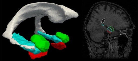
Because the entorhinal cortex memorizes and consolidates this corrupted data pattern across both conscious focal states and recumbent sleep states, attempting to clear the error solely in an upright sitting position leaves the nighttime software patch completely untouched.
Amygdala Threat Tagging and Autonomic Locked States
When the patient disembarks, the physical ground stops moving, but the left entorhinal cortex and the locked brainstem filter continue running the consolidated motion patch, creating an immediate, severe sensory mismatch. The vestibular pathways project directly to the amygdala via the parabrachial nucleus. Recognizing the catastrophic mismatch between expectations of entrained motion and the land's actual physical stillness, the amygdala tags this error as a severe, life-threatening event.
CLINICAL INSIGHT: The Autonomic Superglue. The amygdala’s involvement transforms MdDS from a simple vestibular error into an autonomic emergency. This threat response releases neurochemicals that lock the corrupted software patch in place, ensuring the system remains in a high-stress state that actively resists standard habituation exercises (Mucci et al., 2018).
Otolithic Gravitational Reorientation and Cervical Afferent Chaos
Incorporating recumbent and varied reclining positions into the Zero Out Method is a neurophysiological necessity due to the structural reorientation of the otolith organs relative to gravity. When a patient shifts into a supine or recumbent position, the entire vestibular coordinate frame rotates 90 degrees relative to the constant 1g gravity vector. This rotation flips the functional mechanics of the otoliths: the saccular macula, which normally registers vertical heave, becomes oriented horizontally like a utricle, while the utricular macula rotates to assume a vertical orientation. Additionally, the semicircular canals alter their spatial relationship to gravity, shifting their baseline resting tone.
Critically, this shift cannot be analyzed in isolation from the cervical spine. The deep cervical musculature—specifically the suboccipital complex—provides massive afferent input to the vestibular nuclei regarding head position in space. During long-distance travel, the head is often fixed in a static, strained position for hours, creating an “afferent overload” in which the cervical spine sends conflicting signals that contradict the vestibular and visual systems. The Zero Out Method forces the brain to confront the error while explicitly manipulating the cervical afferent environment, acknowledging that the neck’s proprioceptive input is a primary coordinate in the vestibular map.
Clinical Foundations: The Dai Protocol and the Evolution of MdDS Rehabilitation
The Dai Protocol (Dai et al., 2014) remains a cornerstone of MdDS management, as it was the first clinical methodology to demonstrate that full-field optokinetic stimulation could realign the velocity storage integrator, providing a real pathway to relief. By applying full-field optokinetic stimulation paired with passive head-tilt maneuvers, the Dai Protocol induces significant changes in the vestibulo-ocular reflex, successfully modifying the central processing of motion signals.
The Zero Out Method serves as an evolutionary advancement of this work. While the Dai Protocol established the effectiveness of optokinetic re-alignment, the Zero Out Method optimizes the input to achieve a cleaner, more stable reset by bypassing the nystagmus-driven tracking load. The Zero Out Method avoids the ocular-motor stress of full-field tracking and instead delivers a pure motion signal directly into the subcortical vestibular nuclei. This approach maintains the Dai Protocol’s realignment objective while refining the delivery mechanism to ensure a more stable and efficient clinical outcome.
Advanced Modifications: Mount Sinai and Antwerp Matrix Frameworks
Building upon the clinical success of the Dai Protocol, Dr. Sergei Yakushin at Mount Sinai has significantly advanced this science. Yakushin’s advanced NIH-backed research integrates rotational chair habituation, systematically combining rotary vectors with optokinetic inputs to further isolate and address specific, complex eigenvector deviations that linear protocols miss (Yakushin et al., 2020; Maruta et al., 2024).
Simultaneously, the Antwerp group, led by Dr. Floris Wuyts and Dr. Catho Schoenmaekers, has continuously evolved the clinical standards for MdDS. The Antwerp group’s introduction of the BRSOS (Bilateral Rotational and Somatosensory Stimulation) system, alongside their standardized treatment guidelines and virtual reality trials, demonstrates that cross-modal sensory inputs and highly controlled visual environments are absolutely vital to interrupting the persistent perception of motion (Schoenmaekers et al., 2024).
The Zero Out Method builds directly upon these multi-axial targeting foundations established by Yakushin and Schoenmaekers, but it alters the mechanical visual input by enforcing the foveal wedge.
Key Concept: Rather than exposing the VSI to the potential bottom-up loading from full-field tracking and nystagmus, the stationary central dot completely isolates the peripheral subcortical pathways.
How Does Foveal Suppression Prevent Oculomotor Velocity Loading?
The Zero Out Method completely rejects the bottom-up tracking model and instead introduces a central, stationary focal point to act as an absolute mechanical wedge.

When the patient stares fixedly at this central dot while the visual background moves, the fovea remains totally anchored. This explicit requirement for absolute gaze fixation silences the smooth pursuit system and entirely suppresses the fast-phase resets of optokinetic nystagmus. By shutting down these cortical ocular-motor tracking mechanisms, the clinician prevents any new asymmetric velocity data from being loaded into the velocity storage integrator from the bottom up.
CLINICAL INSIGHT: The Foveal Wedge. By fixing gaze on a central anchor, we shut down the smooth pursuit and saccadic systems. This moves processing from the geniculostriate cortex (the “thinking” brain) directly to the subcortical accessory optic system, delivering a pure motion signal that the brain cannot argue with.
Why Is Top-Down Vection Effective in Reversing MdDS?
This subcortical visual shift allows the clinician to use pure, top-down vection, which is the powerful illusion of passive self-motion, to reverse-engineer the original MdDS onset. Predictive coding models and Neural Mismatch Theory dictate that the central nervous system continuously builds internal models of the physical environment based on past experience (Reason, 1978).
The Zero Out Method gives the patient a form of reverse-engineered adaptation. By generating massive peripheral vection while the eyes remain anchored to a stationary point, the clinician tricks the brain into perceiving the exact same type of passive motion from the top down, mimicking the original linear and angular accelerations. This protocol deliberately reopens the corrupted software file in the central nervous system. Bringing the entrained pattern out of the background allows the nervous system to finally confront, challenge, and rewrite the multi-axis error.
The Role of Primitive Somatosensory-Vestibular Networks in Dynamic Reweighting
To delete this corrupted software patch, the clinician does not attempt to balance the velocity storage integrator with more visual tracking data; instead, the protocol forces the central nervous system to run an error-mitigation check against the absolute physical truth of the environment. Dynamic sensory reweighting relies on the foundational principle that vestibular, proprioceptive, and cervical afferent inputs form the absolute bedrock of postural control and axial stability (Jeka & Anson, 2019). The motor control matrices established by physical therapy researchers dictate that the brain must learn to systematically downweight inaccurate visual data by heavily weighting firm somatosensory contact (Horak & Nashner, 1986; Shumway-Cook & Woollacott, 2012).
The Zero Out Method reactivates this primal processing loop by firmly grounding the patient to a stable surface in various targeted positions, flooding the central nervous system with clean, unyielding mechanoreceptor data from the plantar surfaces, joint proprioceptors, and the cervical spine, paired with the baseline gravity-sensing tone of the otoliths. As modeled by Peterka (2002) in dynamic sensory reweighting frameworks, the brain downweights unreliable visual input and entirely removes the corrupted MdDS software patch (Peterka & Loughlin, 2004). This process re-establishes the fundamental rule of human orientation: “When the ground and the inner ear say I am still, the visual movement is false.”
How Does the Reset Process Degauss the Corrupted Vestibular Cache?
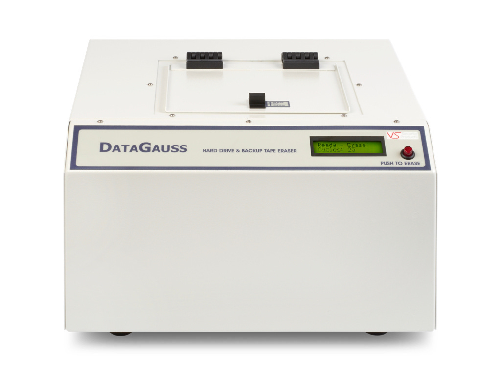
The Zero Out Method fundamentally redefines how clinicians must approach MdDS rehabilitation, shifting the paradigm from gradual adaptation to an immediate systemic reset. Instead of delicately balancing the integrator with visual data, the method acts as a literal degaussing mechanism for the brain’s spatial orientation networks. Degaussing, whether used to protect naval hulls from magnetic mines or to wipe a corrupted hard drive, uses a massive, neutral force to erase a stuck, distorted pattern and reset the system hardware to absolute zero.
CLINICAL INSIGHT: The Degaussing Pulse. The Washout Purge State is the clinical equivalent of a hard drive degausser. By forcing the central nervous system to confront an intense visual illusion while locked to an unyielding physical surface, we create a sensory conflict the brain cannot resolve. It is forced to dump the corrupted data cache to maintain system stability.
The critical bridge between exposing the motion error and deleting the code occurs during the digital engine’s Washout Purge State. If a patient is subjected to continuous optokinetic vection without an explicit window for neural consolidation, the amygdala remains in a state of high alarm. The Washout state breaks this loop by instantly freezing the moving particle field, clearing the visual threat, and replacing it with a prominent, luminous Autonomic Respiratory Pacer (4-7-8 rhythm). This cycle downregulates the sympathetic threat response, providing the “demagnetizing pulse” necessary to break the entrained loop and permanently re-establish the rule of human orientation:
“When the ground says I am still, I am still.”
References
Ayres, A. J. (1972). Sensory Integration and Learning Disorders. Los Angeles, CA: Western Psychological Services.
Ayres, A. J. (1979). Sensory Integration and the Child. Los Angeles, CA: Western Psychological Services.
Brown, J. J., & Baloh, R. W. (1987). Persistent mal de debarquement syndrome: a motion-induced subjective disorder of balance. American Journal of Otolaryngology, 8(4), 219-222. doi: 10.1016/s0196-0709(87)80007-8.
Cha, Y. H. (2009). Mal de debarquement. Seminars in Neurology, 29(5), 520-527. doi: 10.1055/s-0029-1241038.
Cohen, B., Yakushin, S. B., & Cho, C. (2018). Hypothesis: The Vestibular and Cerebellar Basis of the Mal de Debarquement Syndrome. Frontiers in Neurology, 9, 28. doi: 10.3389/fneur.2018.00028.
Dai, M., Cohen, B., Cho, C., Shin, S., & Yakushin, S. B. (2014). Treatment of the Mal de Débarquement Syndrome: a 1-year follow-up. Frontiers in Neurology, 5, 124. doi: 10.3389/fneur.2014.00124.
Darwin, E. (1796). Zoonomia; Or, the Laws of Organic Life, Vol. II. London: J. Johnson.
Hain, T. C., Hanna, P. A., & Rheinberger, M. A. (1999). Mal de debarquement. Archives of Otolaryngology–Head & Neck Surgery, 125(6), 615-620. doi: 10.1001/archotol.125.6.615.
Herdman, S. J., & Clendaniel, R. (2014). Vestibular Rehabilitation (4th ed.). Philadelphia, PA: F.A. Davis Company.
Horak, F. B., & Nashner, L. M. (1986). Central programming of postural movements: adaptation to altered support-surface configurations. Journal of Neurophysiology, 55(6), 1369-1381. doi: 10.1152/jn.1986.55.6.1369.
Irwin, J. A. (1881). The pathology of seasickness. The Lancet, 118(3041), 907-909.
Jeka, J. J., & Anson, E. R. (2019). Sensory reweighting in human postural control. In Mechanisms of Sensory Biology. Springer.
Maruta, J., Cho, C., Raphan, T., & Yakushin, S. B. (2024). Symptom reduction in mal de débarquement syndrome with attenuation of the velocity storage contribution in the central vestibular pathways. Frontiers in Neurology, 15, 1360155. doi: 10.3389/fneur.2024.1360155.
Miller, Michael & Younes, Laurent & Ratnanather, John & Brown, Timothy & Trinh, Huong & Lee, David & Tward, Daniel & Mahon, Pamela & Mori, Susumu & Albert, Marilyn. (2014). Amygdalar Atrophy in Symptomatic AD Based on Diffeomorphometry: The BIOCARD Cohort. Neurobiology of Aging. 36. 10.1016/j.neurobiolaging.2014.06.032.
Mucci, V., Canceri, J. M., Brown, R., Dai, M., Yakushin, S. B., Watson, S. R., & Browne, C. J. (2018). Mal de debarquement syndrome: a survey on subtypes, misdiagnoses, onset and associated psychological features. Journal of Neurology, 265(3), 486-499. doi: 10.1007/s00415-017-8732-2.
Peterka, R. J. (2002). Sensorimotor integration in human postural control. Journal of Neurophysiology, 88(3), 1097-1118. doi: 10.1152/jn.2002.88.3.1097.
Peterka, R. J., & Loughlin, P. J. (2004). Dynamic regulation of sensorimotor integration in human postural control. Journal of Neurophysiology, 91(1), 410-423. doi: 10.1152/jn.00516.2003.
Reason, J. T. (1978). Motion sickness adaptation: a neural mismatch model. Journal of the Royal Society of Medicine, 71(11), 819-829. doi: 10.1177/014107687807101109.
Schoenmaekers, C., Jillings, S., De Laet, C., Zarowski, A., & Wuyts, F. L. (2024). Guideline for standardized approach in the treatment of the Mal de Debarquement syndrome. Frontiers in Neurology, 15, 1359116. doi: 10.3389/fneur.2024.1359116.
Schoenmaekers, C., De Smet, D., Deblieck, C., Van Riel, J., Zarowski, A., & Wuyts, F. L. (2024). A virtual reality application matches the most established treatment for Mal de Debarquement Syndrome: A non-inferiority, randomized, open-label clinical trial. Neurotherapeutics, 21(5), e00390. doi: 10.1016/j.neurot.2024.e00390.
Shumway-Cook, A., & Woollacott, M. H. (2012). Motor Control: Translating Research into Clinical Practice (4th ed.). Philadelphia, PA: Lippincott Williams & Wilkins.
Yakushin, S. B., et al. (2020). Habituation of the velocity storage integrator via rotational chair and optokinetic combinations in persistent MdDS. Frontiers in Neurology.